IMAGE

Fig. S1

ID
ZDB-IMAGE-131022-12
Source
Figures for Johnson et al., 2013
Image
Figure Caption

Fig. S1

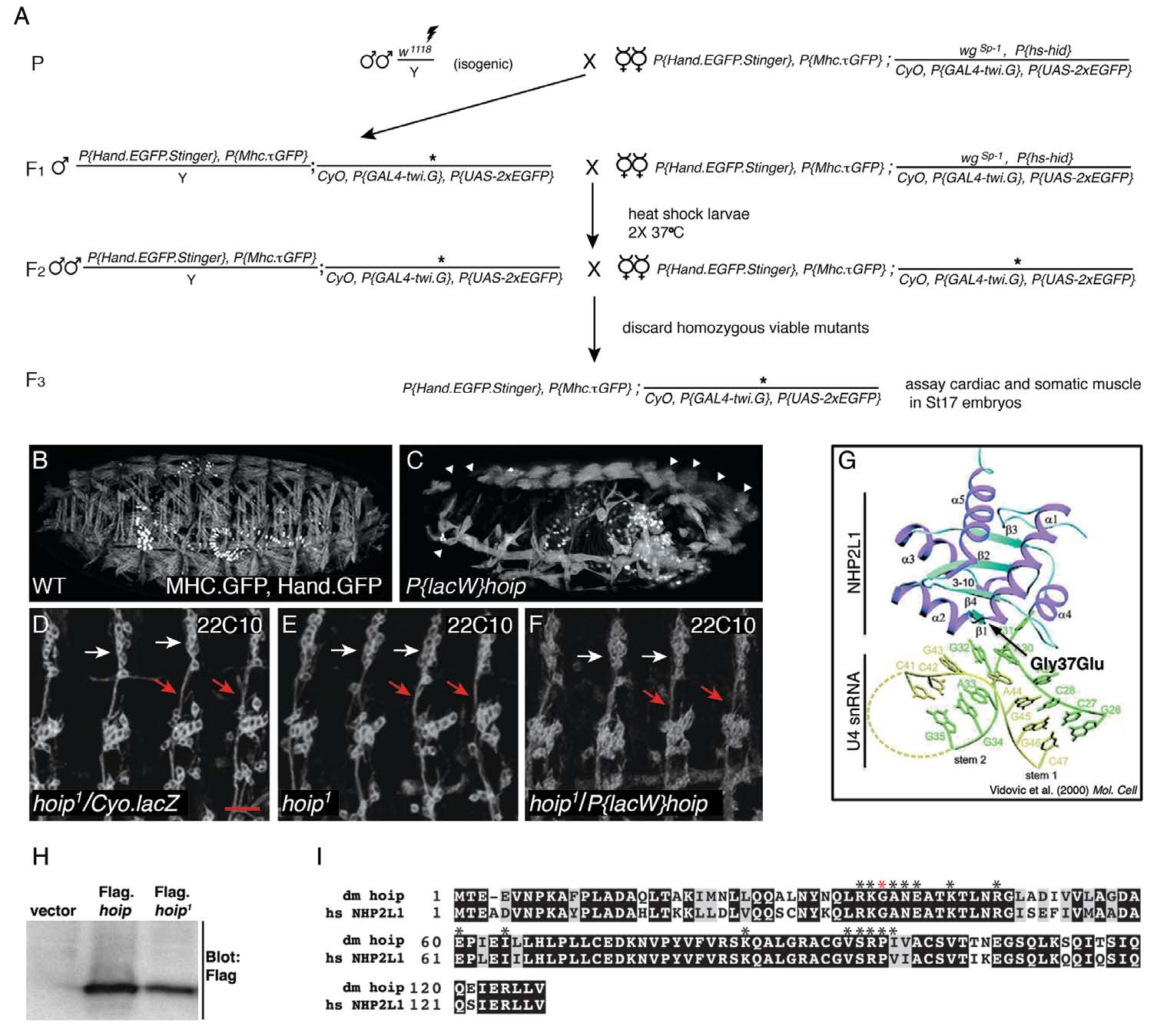
A forward genetic screen identified Hoip as a novel regulator of mesoderm development. (A) Crossing scheme to generate EMS mutants in a double GFP reporter background. Over 10,000 mutagenized genomes were screened. (B,C) MHC.τGFP, Hand.n-GFP expression in St17 embryos. Compared with wild-type embryos (B), P{lacW}hoipk07104 homozygous embryos (C) show severe muscle defects and apparent segmentation defects (white arrowheads). (D-F) St16 embryos stained with the PNS marker 22C10. Micrographs show four dorsal neuron clusters. The organization of the dorsal clusters (white arrows) and the pathway of the descending nerve (red arrows) are comparable among hoip1/Cyo (D), hoip1 (hoip1/P{lacW}hoipk07104 (F) embryos. (G) Crystal structure of hNHP2L1 bound to U4 snRNA as reported previously (Vidovic et al., 2000), except the position of the 37th residue (mutated in hoip1 embryos) is shown. (H) α-Flag western blot from COS cells transfected with empty pcDNA.Flag, pcDNA.Flag.Hoip or pcDNA.Flag.Hoip1. (I) Alignment of Hoip and hNHP2L1 protein sequence. The two proteins are 79% identical and 89% similar. hNHP2L1 amino acids required for RNA binding are indicated with an asterisk (Schultz et al., 2006). The red asterisk shows the position of the hoip1 missense mutation. Scale bar: 20μm.

Acknowledgments
This image is the copyrighted work of the attributed author or publisher, and ZFIN has permission only to display this image to its users. Additional permissions should be obtained from the applicable author or publisher of the image. Full text @ Development