
|
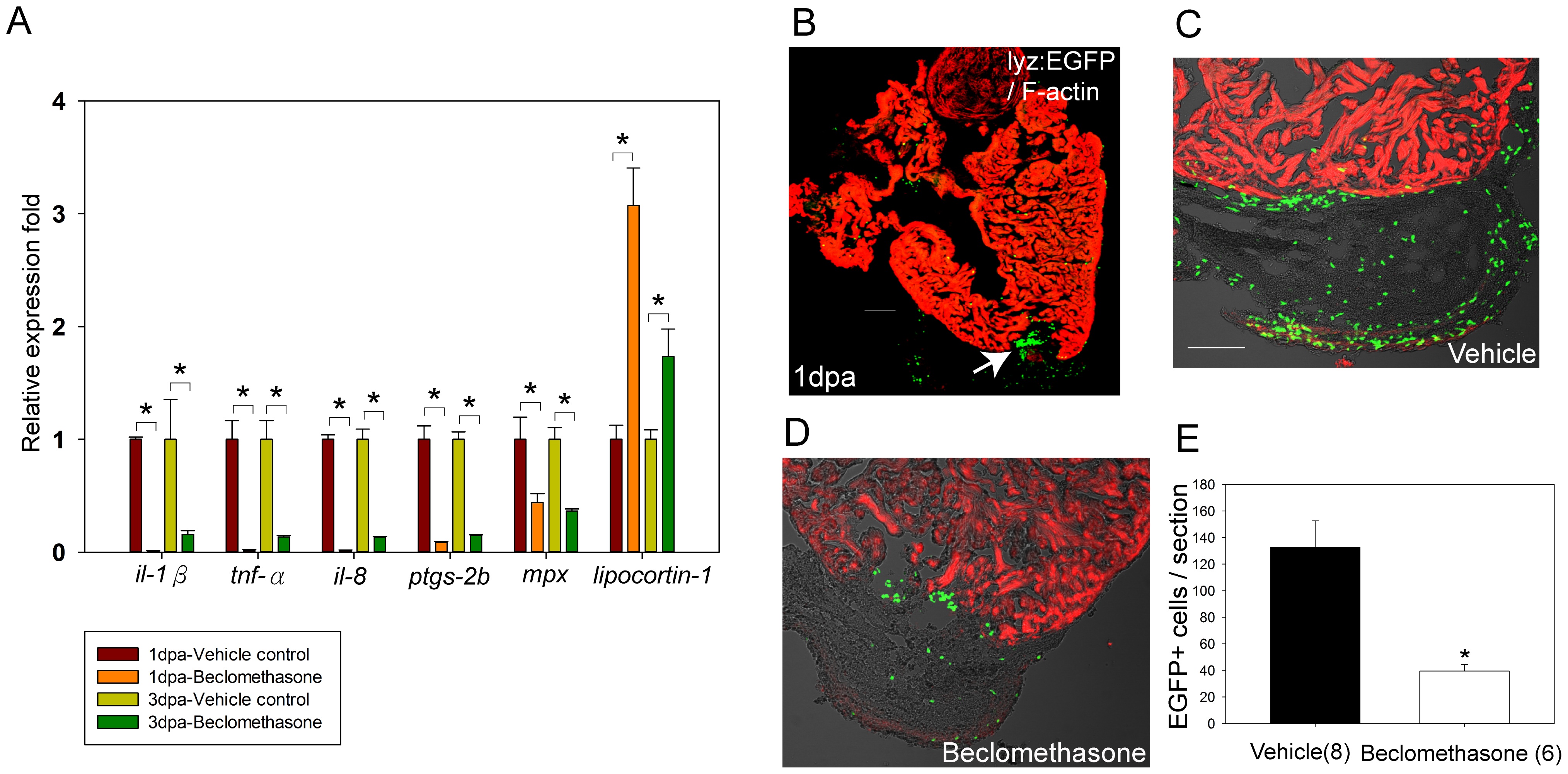
Fig. 3
Beclomethasone treatment reduced pro-inflammatory gene expression and phagocyte recruitment after cardiac injury.
Whole hearts were harvested at 1 day and 3 days post ventricular resection. RT-qPCR was conducted to quantify the relative fold of pro-inflammatory genes at hearts. (A) Expression of il-1β, tnf-α, il-8, ptgs-2b, and mpx were all significantly reduced in the beclomethasone-treated group for at least 3 days, while lipocortin-1 expression was significantly induced after beclomethasone treatment (n = 3). * indicates p<0.05. (B) Immunostaining of hearts after injury. Green: phagocytes (lyz-EGFP); red: Phalloidin-546. After cardiac injury, numerous phagocytes accumulated in the wound (arrow). (C, E) In the vehicle control group, average of 133±20 phagocytic cells were counted in the fibrin clots (n = 8). (D, E) In the beclomethasone treated animals, 40±5 cells were counted (n = 6). (E) Treatment with beclomethasone significantly impaired phagocyte recruitment in the injured hearts. The data represent the mean±SEM, *indicates p<0.05, scale bar = 100 μm.