
|
Fig. 6
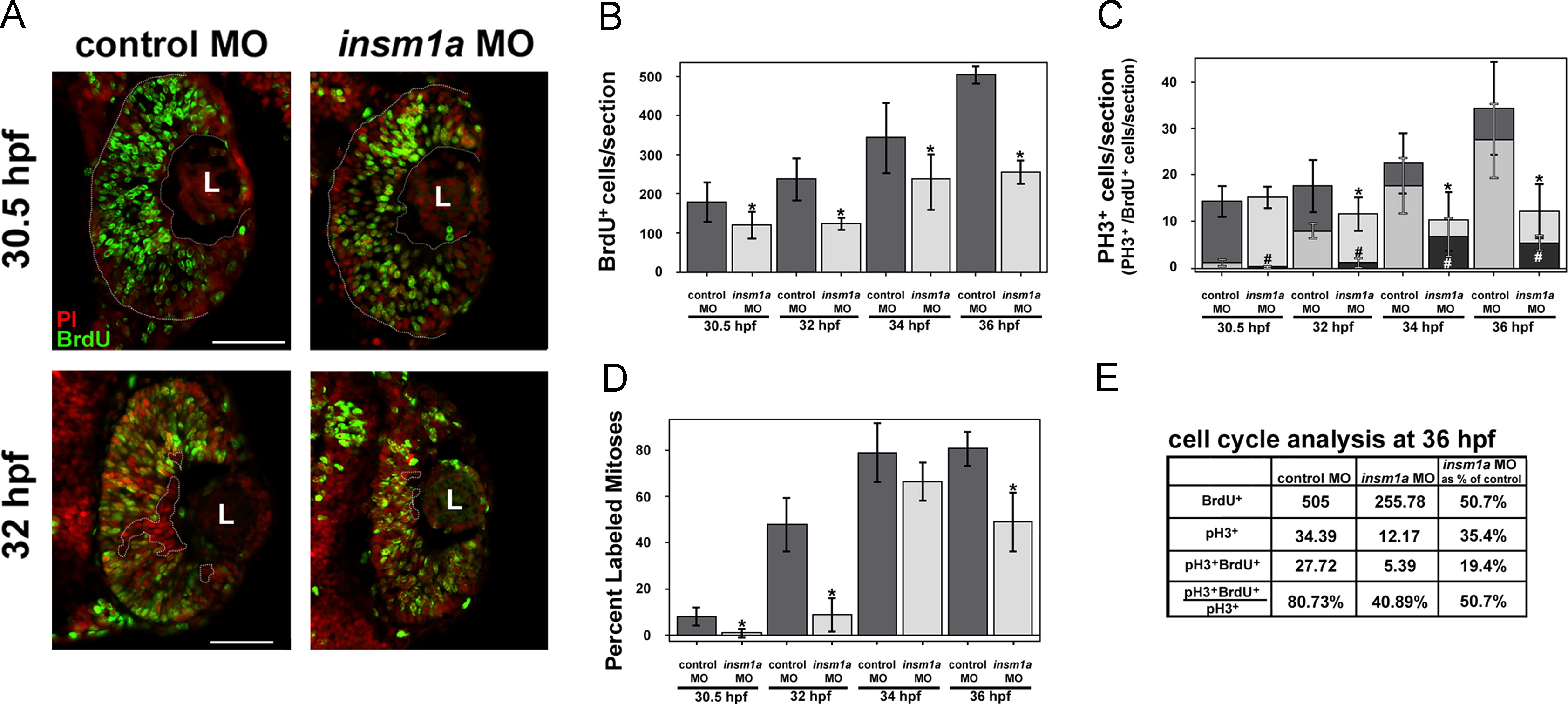
Cell cycle progression is delayed after insm1a knockdown. (A) At 30.5 hpf (0.5 hpi, top panels), BrdU+ cells (green) spanned the retina from basal to apical (dotted lines) in control retinas. However, in insm1a morphant retinas, the BrdU+ cells did not extend to the apical edge of the retina. At 32 hpf (2 hpi, bottom panels), fewer post-mitotic BrdU- cells were observed at the basal surface (dotted circles) in insm1a morphants compared with controls, indicating a delay in cell cycle exit in insm1a morphant retinas. (B) Quantitation of BrdU+ cells revealed a decrease in insm1a morphant retinas at all time points (*p<0.05). (C) The numbers of PH3+ cells were reduced at 2, 4 and 6 hpi in insm1a morphant retinas (*p<0.04). The number of cells double-positive for PH3 and BrdU was also reduced at all time points (#p<0.007). (D) The percent labeled mitoses (see Results for explanation) was significantly reduced in insm1a morphants at 0.5, 2 and 6 hpi, indicating that insm1a morphants took longer to progress from S phase into late G2/M phase compared with controls. (E) Summary of cell count analysis at 36 hpf (6 hpi); cell counts for insm1a morphants are expressed as a percentage of controls in the last column (for both controls and morphants: n=6 at 30.5, 32, and 34 hpf and n=3 at 36 hpf; mean±st.dev). Scale bars=50 μm; L, lens; PI, propidium iodide; hpf, hours post fertilization; hpi, hours post BrdU injection; MO, morpholino.
Reprinted from Developmental Biology, 380(2), Forbes-Osborne, M.A., Wilson, S.G., and Morris, A.C., Insulinoma-associated 1a (Insm1a) is required for photoreceptor differentiation in the zebrafish retina, 157-171, Copyright (2013) with permission from Elsevier. Full text @ Dev. Biol.