
Image
|
Figure Caption
Fig. S2
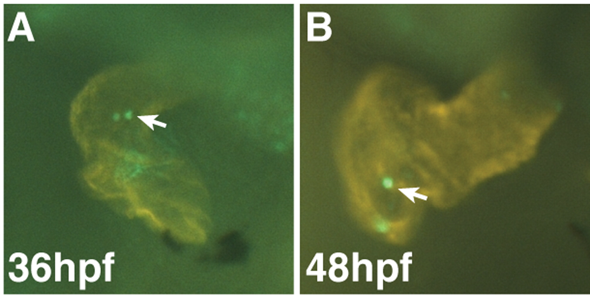
Representative images of hearts with pHH3+ CMs. (A) Representative WT heart at 36 hpf. (B) Representative WT heart at 48 hpf. MF20 (red) was used to label CMs. pHH3+ cells (green; arrows) were easily discernible in the CMs and distinct from non-CMs when using the fluorescent stereomicroscope focusing through the heart.
Acknowledgments
This image is the copyrighted work of the attributed author or publisher, and
ZFIN has permission only to display this image to its users.
Additional permissions should be obtained from the applicable author or publisher of the image.
Reprinted from Developmental Biology, 380(2), Sorrell, M.R., Dohn, T.E., D'Aniello, E., and Waxman, J.S., Tcf7l1 proteins cell autonomously restrict cardiomyocyte and promote endothelial specification in zebrafish, 199-210, Copyright (2013) with permission from Elsevier. Full text @ Dev. Biol.