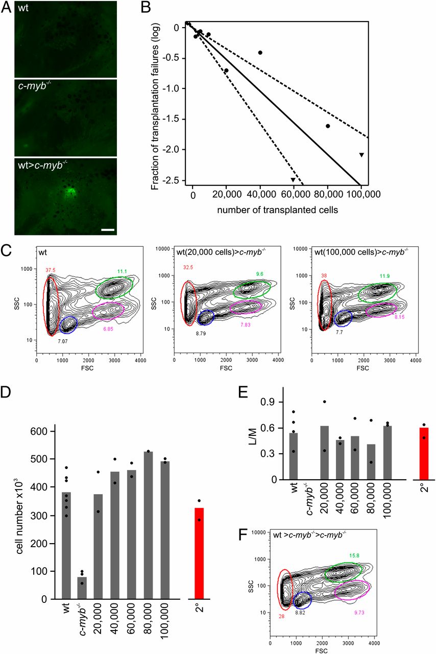
Fig. 3 Long-term multilineage reconstitution of c-myb-/- mutant fish. (A) Thymus colonization in c-myb-/- recipients transplanted with wild-type ikaros:eGFP-transgenic WKM cells. Photographs (side views) were taken 7 d after transplantation. (Scale bar, 50 μm.) (B) Limiting dilution analysis of hematopoietic reconstitution of c-myb-/- mutant fish. (C) Flow cytometric analysis of WKM of wild-type fish (Left) and stably reconstituted c-myb-/- mutants after transplantation of 20,000 (Middle) and 100,000 (Right) wild-type WKM cells (see Fig. 1E legend for explanation of indicated cell populations). (D) Cellularity of WKM of wild-type (wt) fish, c-myb-/- mutants, and mutants stably reconstituted after transfer of the indicated numbers of WKM wild-type cells (9 wk after transplantation). The mean values (bars) and results of individual fish are shown. The number of cells found in WKM in secondary transplant recipients (2°) is depicted in red. (E) Multilineage reconstitution as measured by the ratio of cells in lymphoid (L) and myeloid (M) gates in flow cytometric analyses for fish depicted in D. (F) Flow cytometric analysis of WKM of stably reconstituted c-myb-/- mutants after transplantation of WKM of primary recipients (see Fig. 1E legend for explanation of indicated cell populations).
Image
Figure Caption
Acknowledgments
This image is the copyrighted work of the attributed author or publisher, and
ZFIN has permission only to display this image to its users.
Additional permissions should be obtained from the applicable author or publisher of the image.
Full text @ Proc. Natl. Acad. Sci. USA

