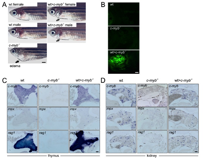
Fig. S1 Characterization of reconstituted c-myb-/- mutants. (A) Side views of wild-type, c-myb-/- transplant recipients and a c-myb-/- mutant fish. Note the absence of cardiac edema (resulting in a bulged body curvature) in the transplanted fish; the body curvature is highlighted by dotted lines. (Scale bar, 1 mm.) (B) Reconstitution of T-cell development in the thymus of c-myb-/- mutant recipients transplanted with ikaros:eGFP-transgenic wild-type whole kidney marrow (WKM) cells; note the presence of green fluorescent cells in the transplanted fish. Photographs (side views) were taken 20 d after transplantation. (Scale bar, 50 μm.) (C and D) Whole-mount RNA in situ hybridization with c-myb-, mpx-, and rag-specific probes on thymus (C) and kidney (D) sections of wild-type, c-myb-/- mutant and c-myb-/- transplant recipients (3 wk after transplantation). In mutant tissues, no positive cells were observed for c-myb, mpx, and rag1, while expression patterns in transplanted c-myb-/- recipients are indistinguishable from those of their wild-type siblings. c-myb is a general marker for hematopoietic cells; mpx is a marker for myeloid cells; rag1 is a marker for developing lymphocytes. (Scale bar, 50 μm.)
Image
Figure Caption
Figure Data
Acknowledgments
This image is the copyrighted work of the attributed author or publisher, and
ZFIN has permission only to display this image to its users.
Additional permissions should be obtained from the applicable author or publisher of the image.
Full text @ Proc. Natl. Acad. Sci. USA

