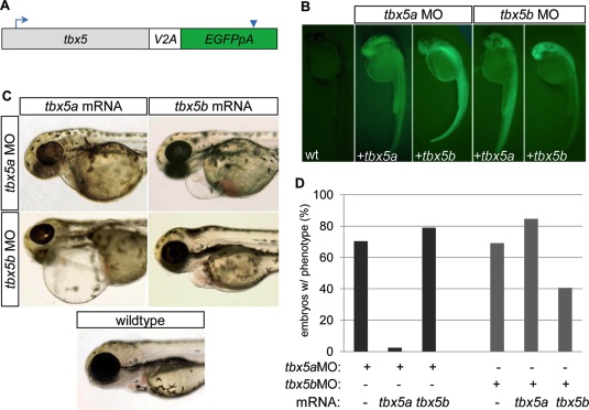
Fig. 10 mRNA injection rescue studies indicate that tbx5a and tbx5b are not functionally redundant in cardiac development. A: Schematic of mRNA structure for rescue constructs, in which the coding sequence for tbx5a or tbx5b was connected to enhanced green fluorescent protein (EGFP, with accompanying poly-adenylation signal) by means of a viral 2A peptide. Blue arrow indicates location of translation start site. Blue arrowhead indicates termination signal. B: Fluorescent images of 26 hours postfertilization (hpf) embryos previously injected with combinations of either buffer (in wild-type, wt), tbx5aMO or tbx5bMO, and tbx5a or tbx5b mRNA to evaluate mRNA integrity by means of production of translated GFP. C: Brightfield images of 72 hpf embryos showing representative cardiac phenotypes for each group of injected embryos. D: Quantification of morpholino injected embryos displaying a cardiac morphology phenotype at 54 hpf. (tbx5aMO 70.3% n = 91; tbx5a MO+ tbx5a mRNA 2.4% n = 84; tbx5a MO+ tbx5b mRNA 78.9% n = 133; tbx5b MO 69.1% n = 220; tbx5b MO+ tbx5a mRNA 84.6% n = 65, tbx5b MO+ tbx5b mRNA 40.6% n = 69).
Image
Figure Caption
Figure Data
Acknowledgments
This image is the copyrighted work of the attributed author or publisher, and
ZFIN has permission only to display this image to its users.
Additional permissions should be obtained from the applicable author or publisher of the image.
Full text @ Dev. Dyn.

