
|
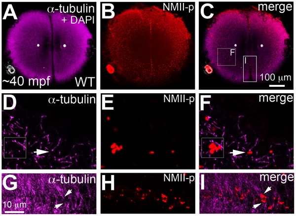
Fig. 6 Germ plasm RNPs localize to the tips of astral microtubules.
Animal views of blastodisc cortex. (A–C) GP RNPs are located in a band (B) at the cortical periphery. Higher magnification images indicate that multimerized GP RNPs are located at the tips of spindle pole astral microtubules (D–F; G–I). In non-furrow peripheral regions (D–F), several microtubule tips (D) can be observed to coincide with a large multimerized GP RNP (E, F). Arrow in F points to a smaller multimerized aggregate at a microtubule tip. At the cleavage furrow (G–I), multimerized GP RNPs are found as bilateral rows of aggregates (arrows in I) at bundled tips of astral microtubules from contralateral sides (G–I). Panels G–I are high magnification views of the rectangular box (labeled I) in panel C, rotated 90° counterclockwise. Panels D–F are high magnification views from an embryo comparable to that in C of a non-furrow region similar to the square box indicated in panel C (labeled F).