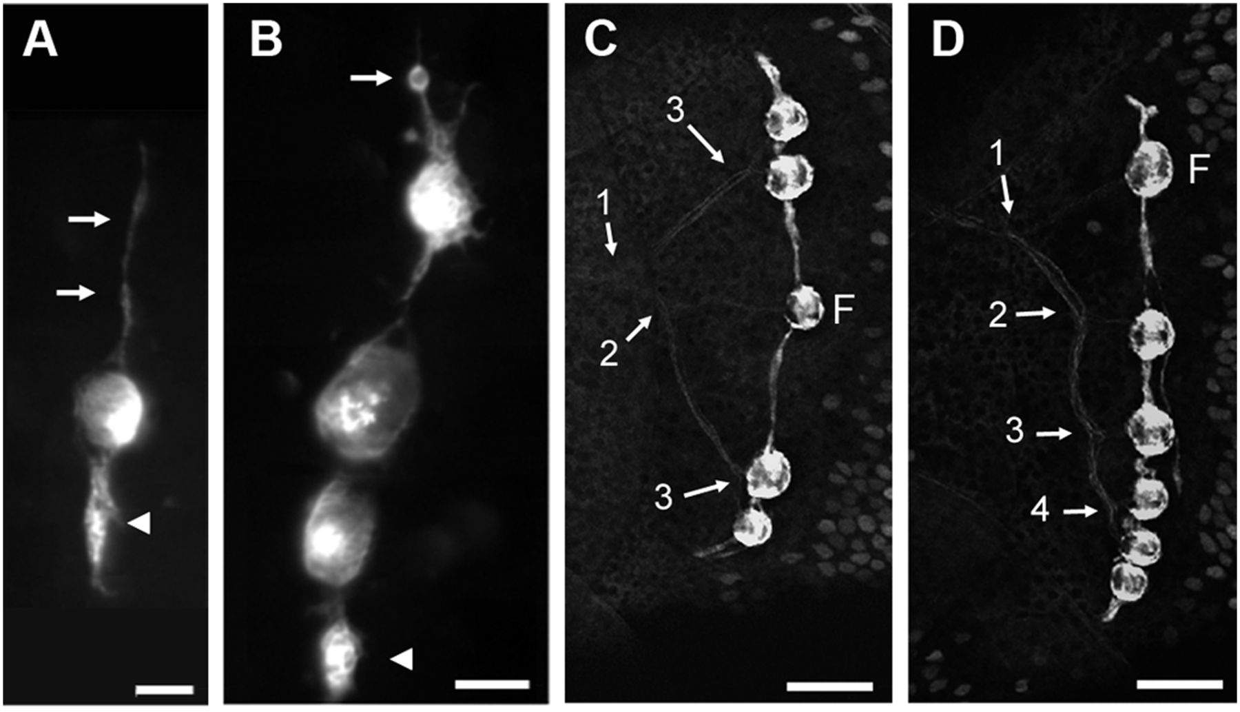
Fig. 1 Stitches in zebrafish. (A and B) Initiation of stitch formation in cxcr4.139:rfp fish. Long cell processes extend from the founder neuromast along the dorsoventral axis (A, arrows) and progressively swell up (arrowhead). Cell proliferation (B, arrow: cell rounding up before mitosis) eventually leads to the formation of a new, complete accessory neuromasts (B, arrowhead). (C and D) Innervation of stitches in Hgn39d; ET20 fish. The branching pattern reveals the history of stitch formation. Numbers refer to the successive branching/budding events. F: founder neuromast. In all figures, anterior is left and dorsal is up. (Scale bars: A and B—20 μm; C and D—50 μm.)
Image
Figure Caption
Figure Data
Acknowledgments
This image is the copyrighted work of the attributed author or publisher, and
ZFIN has permission only to display this image to its users.
Additional permissions should be obtained from the applicable author or publisher of the image.
Full text @ Proc. Natl. Acad. Sci. USA

