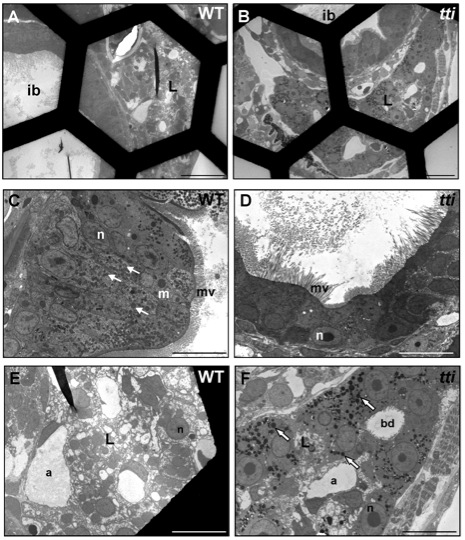
Fig. S6 Absence of dead cells in the intestinal lumen of WT and ttis450 larvae at 7 dpf. (A–F) Transmission electron micrographs of transverse sections of WT and ttis450 larvae at 168 hpf (7 dpf). The number of conspicuous autophagosome-like structures in the IECs of ttis450 larvae has diminished by 7 dpf and there are no dead cells in the lumen (D). Meanwhile, liver cells of ttis450 larvae contain abundant autolysosome-like structures at this time-point (F, white arrows). Scale bars = 50 μm (A, B); 10 µm (C–F). ib, intestinal bulb; n, nucleus; m, mitochondria; mv, microvilli; l, liver; bd, bile duct; a, arteriole.
Image
Figure Caption
Acknowledgments
This image is the copyrighted work of the attributed author or publisher, and
ZFIN has permission only to display this image to its users.
Additional permissions should be obtained from the applicable author or publisher of the image.
Full text @ PLoS Genet.

