Image
Figure Caption
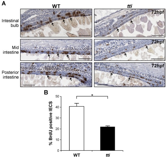
Fig. S1 ttis450 larvae contain fewer replicating IECs than WT larvae. (A) Sagittal sections of the intestine of WT and ttis450 zebrafish larvae at 72 hpf showing cells that accumulated BrdU (black arrows) during a 30 min exposure to this thymidine analogue at 72 hpf. BrdU-positive nuclei (brown) indicate cells in the S-phase of the cell cycle. Scale bars = 50 μm. (B) Quantitation of BrdU-positive IECs in three independent sagittal sections of WT and ttis450 larvae at 72 hpf reveals that ttis450 larvae contain approximately 50% fewer S-phase IECs than WT. *p<0.05. Data are represented as mean +/ SD.
Acknowledgments
This image is the copyrighted work of the attributed author or publisher, and
ZFIN has permission only to display this image to its users.
Additional permissions should be obtained from the applicable author or publisher of the image.
Full text @ PLoS Genet.

