Fig. 4
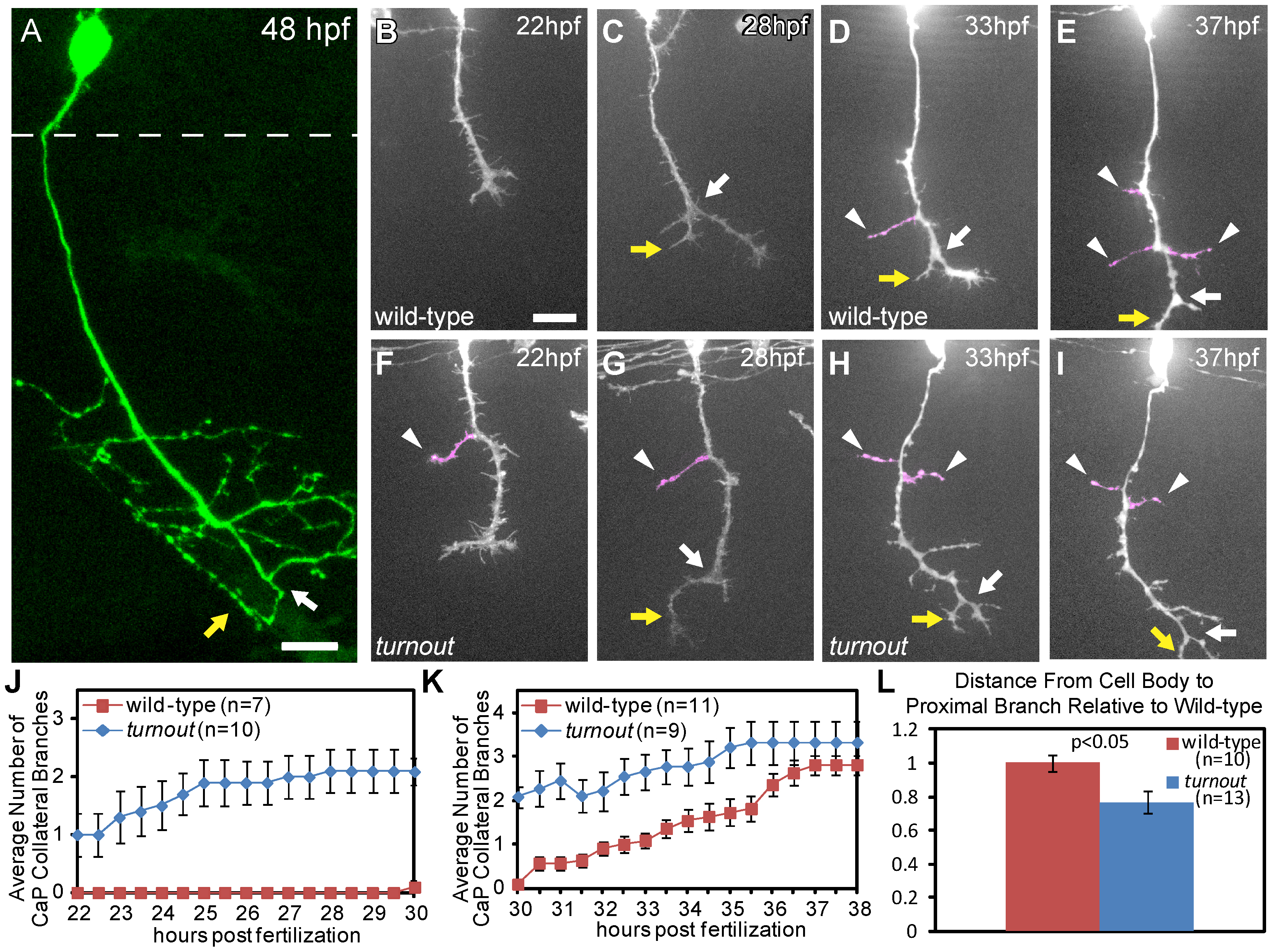
The turnout gene controls where and when collateral CaP axon branches form.
(A) 48 hpf wild-type embryo with a single mnx1:GFP positive CaP motor axon. Note that the axon is devoid of branches along the proximal axon shaft, and that collateral branches only form along the distal axon segment, adjacent to the bifurcation branch (white arrow) with one branch turning rostrally (yellow arrow) and one caudally. Dashed line indicates spinal cord boundary. (B, C) Time-lapse of wild-type CaP axons traversing the ventral myotome between 22 hpf and 30 hpf. (D, E) In wild-type embryos, branching begins at ∼30 hpf, typically by axon bifurcation (arrow), followed by the formation of axon collateral branches (arrowhead). (F, G) turnout CaP axons form precocious collateral branches (arrowhead). (H, I) turnout axon collateral branching plateaus between 30 hpf and 38 hpf. (J) Quantification of CaP axon branches between 22–30 hpf. (K) Quantification of CaP axon branches between 30–38 hpf. (L) Quantification of the location of the first proximal collateral relative to wild-type at 48 hpf. Branches are defined as protrusions longer than 5 µm that persist for longer than 2 hours. Error bars indicate SEM. Scale bar, 15 µm.

