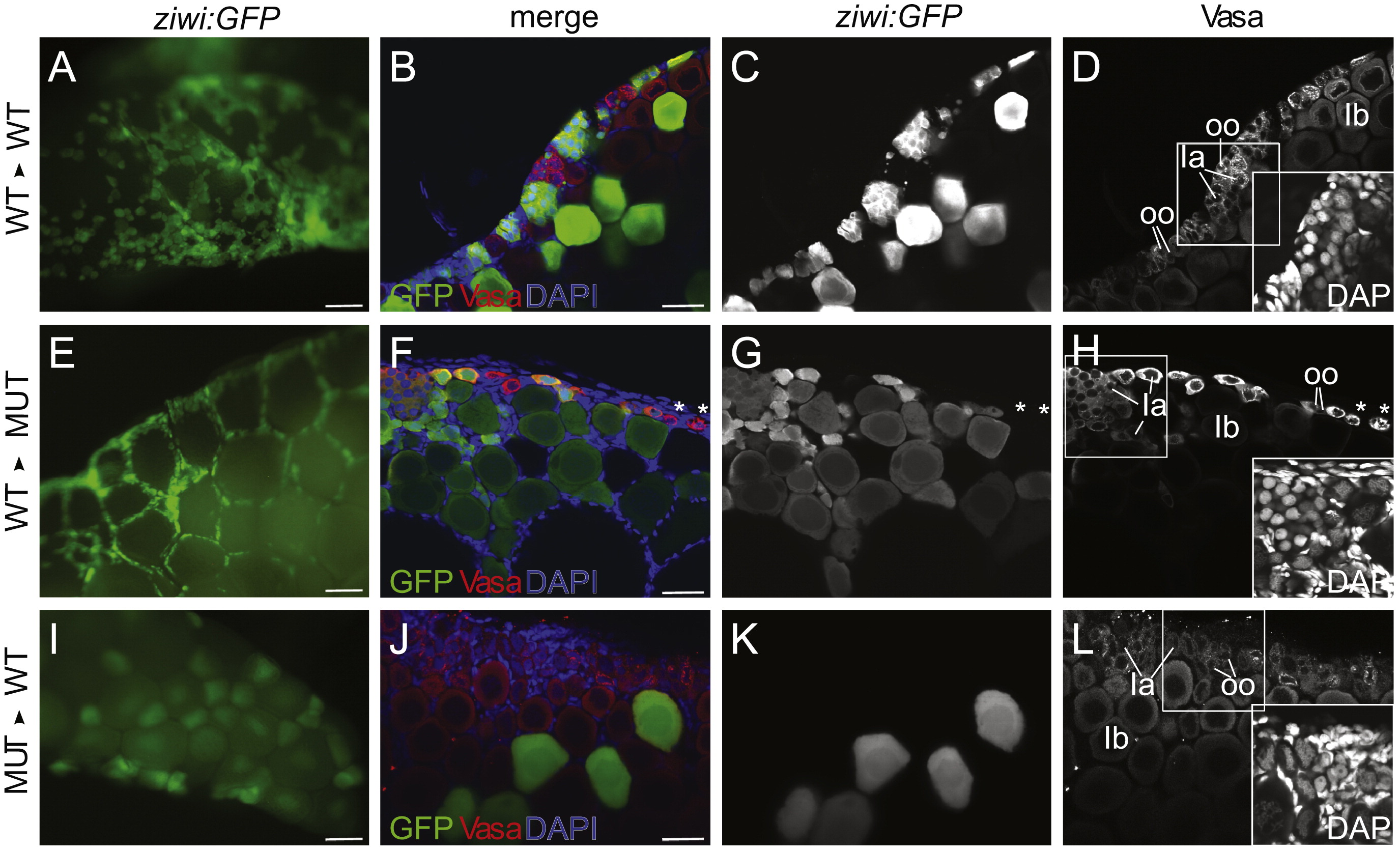
Fig. 5
nanos3 is required cell-autonomously in the <20 μm germ cell population. (A–L) Genetic germline chimeras were created by transplanting Tg(ziwi:EGFP) donor germ cells into EGFP-negative hosts, and the contribution of EGFP+ germ cells was analyzed at 60 dpf. (A, E and I) Low magnification images, where donor derived cells are green. All others panels are higher magnification confocal images. (A–D) In controls, wild-type Tg(ziwi:EGFP) germ cells were able to equally contribute to the <20 μm germ cell population as wild-type host germ cells (WT′WT, n=10). (E-H) Wild-type Tg(ziwi:EGFP) germ cells are the only <20 μm germ cells detected in a nanos3 mutant host ovary (WT′MUT, n=5). (I–L) Conversely, nanos3 mutant; Tg(ziwi:EGFP) donor germ cells are not able to contribute to the <20 μm germ cell population in wild-type host ovaries (MUT′WT, n=2). B, F and J show merged images of EGFP expressing donor cells (green), Vasa expressing germ cells (red), and DNA (blue). C, G, and K, show endogenous GFP expression. D, H and L show Vasa protein localization, and inset shows the DNA morphology of DAPI stained cells boxed in main image. All images are from whole mount preparations. Scale bars: 200 μm (A, E and I), 20 μm (B, F and J). Stage Ia oocytes (Ia), stage Ib oocytes (Ib) and oogonia (oo).
Reprinted from Developmental Biology, 374(2), Beer, R.L., and Draper, B.W., nanos3 maintains germline stem cells and expression of the conserved germline stem cell gene nanos2 in the zebrafish ovary, 308-318, Copyright (2013) with permission from Elsevier. Full text @ Dev. Biol.

