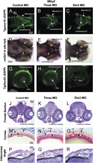
Fig. 2 TH is required for craniofacial and ocular development. 96 hpf control embryos demonstrated neural crest-derived structures including 7 PAs (Meckels cartilage (1st PA), ceratohyal cartilage (2nd PA), and 5 posterior PAs) in vivo in Tg(sox10::EGFP) zebrafish (A) and by col2a1 in situ hybridization (D). Control embryos also showed foxd3 expression in photoreceptors at 96 hpf Tg(foxd3::EGFP) embryos (G). Thraa MO knockdown inhibited Meckels and ceratohyal cartilage formation, decreased the number of posterior PAs (B, E), and decreased photoreceptor expression of foxd3 (H). Dio3 MO knockdown inhibited posterior PA formation (C, F) and foxd3 expression in photoreceptors (I). Dio3 MO knockdown had less effect on Meckels and ceratohyal cartilage formation. Methylacrylate sections demonstrated that control embryos (J) had corneas (M) that contained epithelial (C Epi), stromal (C S), and endothelial (C End) layers, lens epithelial cells (Lens Epi) adjacent to the cornea, and iris stroma with xanthophores (Xn), iridophores (Ir), and undifferentiated cells (Un). Thraa MO knockdowns (K, N, Q, T) demonstrated thickened and scalloped C Epi, lack of C End (N) and decreased cellularity of the iridocorneal angle (Q) vs. control (J, M, P). ON, optic nerve; MR, medial rectus. Scale bar=5, 10, 50 or 100 µm as indicated.
Reprinted from Developmental Biology, 373(2), Bohnsack, B.L., and Kahana, A., Thyroid hormone and retinoic acid interact to regulate zebrafish craniofacial neural crest development, 300-309, Copyright (2013) with permission from Elsevier. Full text @ Dev. Biol.

