Image
Figure Caption
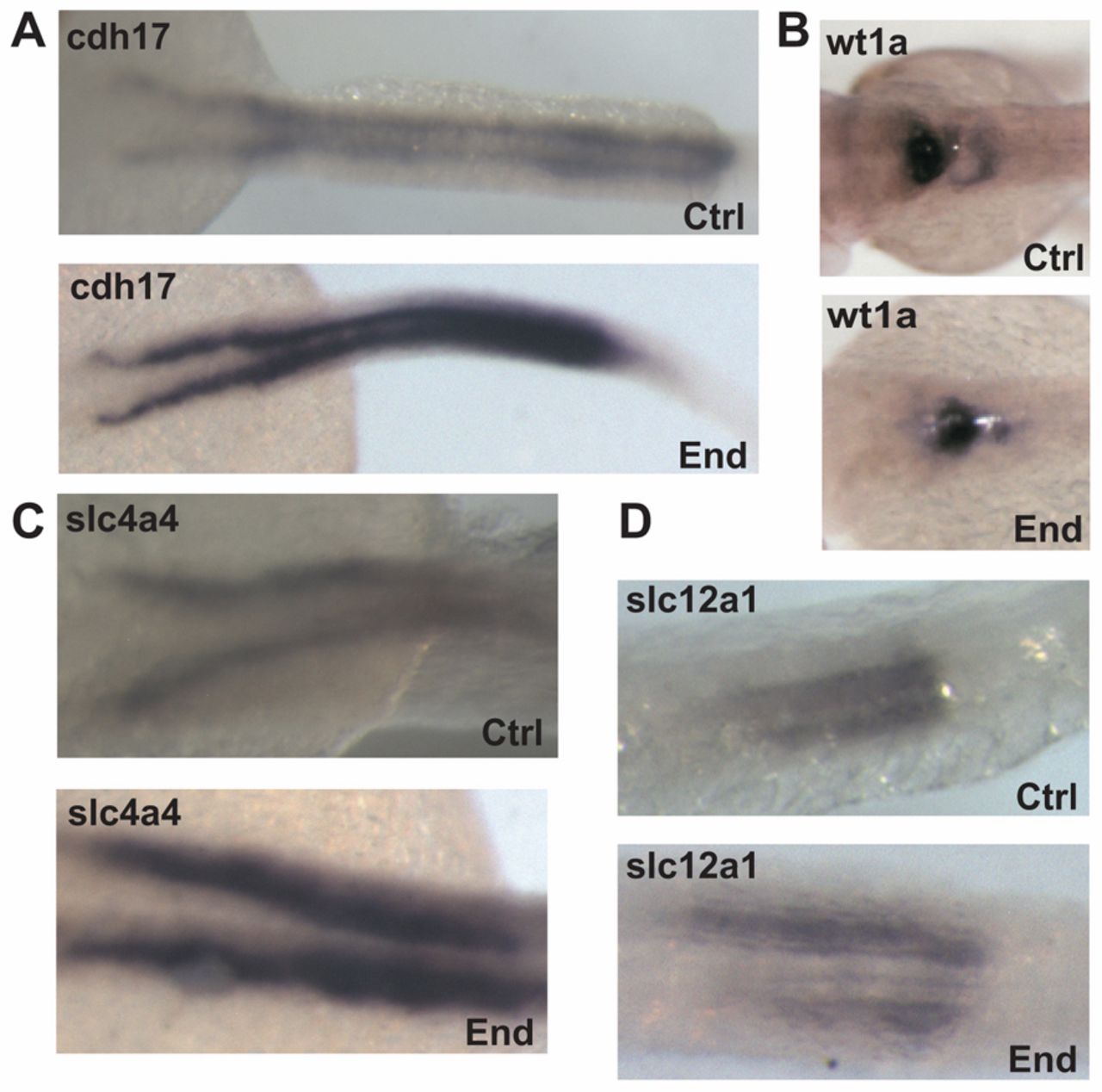
Fig. 6 Kidney morphology is intact after endolyn knockdown. (A) In situ hybridization was performed at 48 hpf to detect cdh17 expression in embryos injected with either control (Ctrl) or endolyn (End) MOs. (B-D) In situ hybridization was performed at 48 hpf in embryos injected with either control or endolyn MO to detect markers for podocytes (wt1a) (B), proximal tubule (slc4a4) (C), and distal tubule (slc12a1) (D). The morphology of the pronephric kidney in morphants is not grossly disrupted, although the tubules appear dilated. Class I morphants were used in all experiments.
Figure Data
Acknowledgments
This image is the copyrighted work of the attributed author or publisher, and
ZFIN has permission only to display this image to its users.
Additional permissions should be obtained from the applicable author or publisher of the image.
Full text @ J. Cell Sci.

