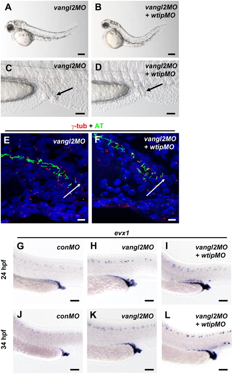
Fig. 5 In vangl2 knockdown embryos, cloaca malformation is associated with fewer ciliated cells.
(A,B) Side view of embryos at 48 hpf imaged with light microscopy. (C,D) Lateral view of cloaca at 48 hpf. The black arrow marks the cloaca. 48 hpf vangl2 (A,C) and vangl2 + wtip (B,D) morphants form pronephric cysts, cloaca malformation, hydrocephalus, body curvature and pericardial edema. Confocal projections of a double immunofluorescence of the posterior pronephros and cloaca in 24 hpf vangl2 (E) and vangl2 + wtip morphant (F) embryos labeled with anti-γ-tubulin (basal bodies, red) and anti-acetylated α-tubulin (cilia, green) revealed fewer ciliated cells in the vangl2 (E) and vangl2 + wtip morphants (F). The white arrow points to the cloaca area. Lateral views of evx1 expression in control (G,J), vangl2 (H,K) and vangl2 + wtip morphants (I,L) in the posterior pronephros and cloaca at 24hpf (G–I) and 34 hpf (J–L). In 24 hpf (G) and 34 hpf (J) control embryos, evx1 was expressed in the epidermis and cloaca. In vangl2, and vangl2 + wtip morphants evx1 was expressed broadly around the area of the posterior pronephros and cloaca at 24 hpf (H,I). evx1 continued to be expressed at 34 hpf in the malformed cloaca (K,L). Scale bars are 200 μm in A,B, 50 μm in C,D,G–L, and 10 μm in E,F.

