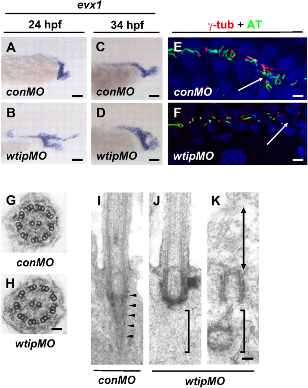
Fig. 4 In wtip knockdown embryos cloaca malformation is associated with fewer ciliated cells and the lack of rootlet structures, which may compromise the motility of cilia.
Lateral views of evx1 expression in control (A,C) and wtip morphants (B,D) in the posterior pronephros and cloaca at 24hpf (A,B) and 34 hpf (C,D). In 24 hpf (A) and 34 hpf (C) control embryos, evx1 was expressed in the epidermis and cloaca. In wtip morphants, evx1 was expressed broadly around the area of the posterior pronephros and cloaca at 24 hpf (B). evx1 continued to be expressed at 34 hpf in the malformed cloaca (D). Confocal projections of a double immunofluorescence of the posterior pronephros and cloaca in 24 hpf control (E) and wtip morphant (F) embryos labeled with anti-γ-tubulin (basal body, red) and anti-acetylated α-tubulin (cilia, green) revealed fewer ciliated cell in the wtip morphants (F). The white arrow points to the cloaca area. Transmission electron micrographs of the transverse sections in 34 hpf control embryos (G) and wtip morphants (H) revealed an axoneme with 9+2 structure. Longitudinal sections in control embryos displayed a striated rootlet structure (arrowheads) located adjacent to the basal body (I). In wtip morphants, ciliated cells exhibited a basal body but lacked rootlet structure (J), and non-ciliated (double arrow) cells exhibited mother centrioles/basal bodies, which were separated from the inner leaflet of the apical cell membrane (K). Scale bars are 25 μm in A–D, 100 μm in E,F, 50 nm in G,H, and 100 nm in I–K.

