Image
Figure Caption
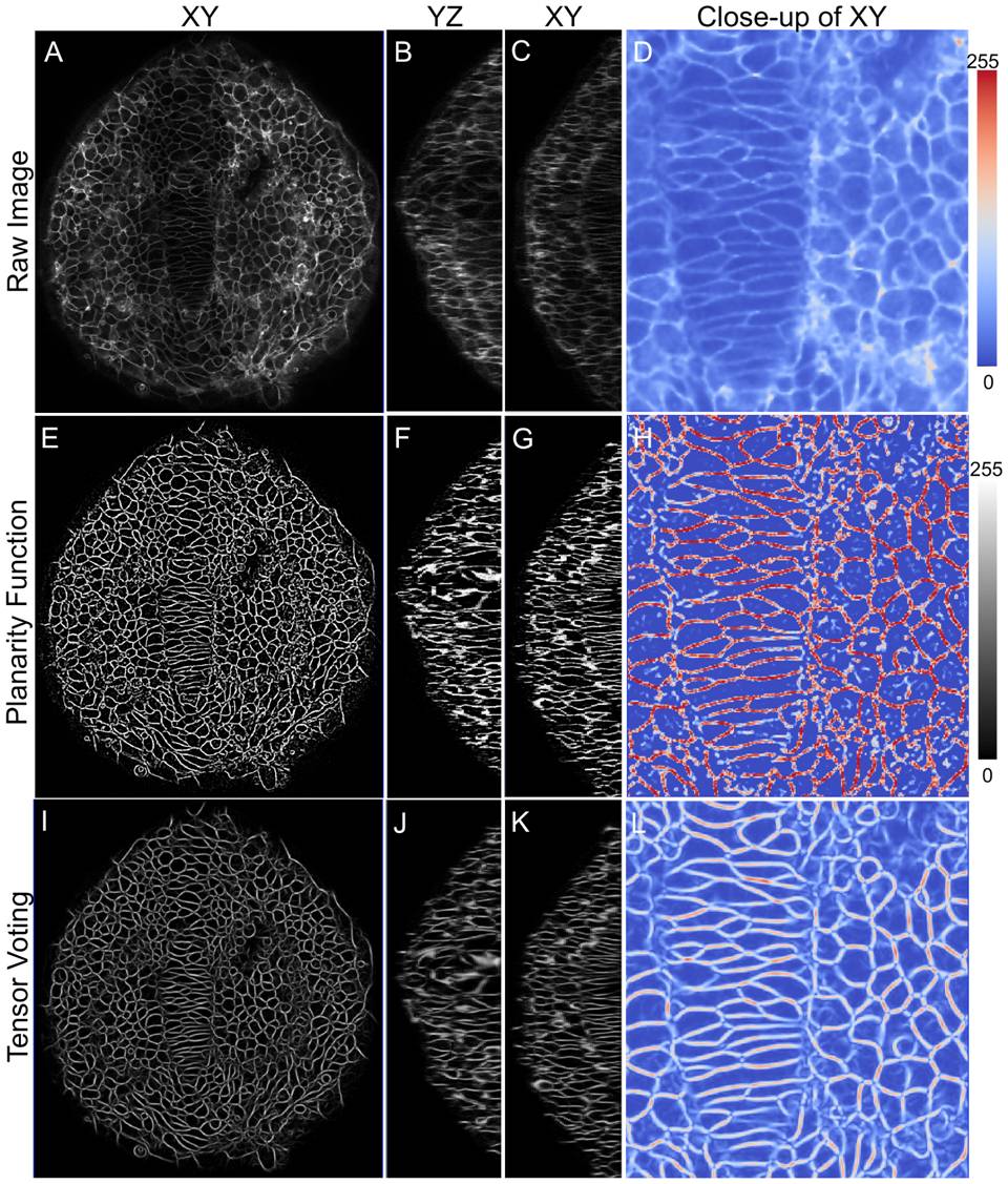
Fig. 4 High-fidelity reconstruction of zebrafish membrane images.
Significant improvement in membrane signal quality is shown in XY, XZ and YZ planes. (A-D) Raw data showing dorsal view (anterior on top) of zebrafish neuroepithelium (ne) and notochord at 12 hpf, (E-H) Planarity function intermediate output and (I-L) Tensor voting final output. The last image in each panel shows a color-mapped zoomed view for easy comparison.
Acknowledgments
This image is the copyrighted work of the attributed author or publisher, and
ZFIN has permission only to display this image to its users.
Additional permissions should be obtained from the applicable author or publisher of the image.
Full text @ PLoS Comput. Biol.

