
|
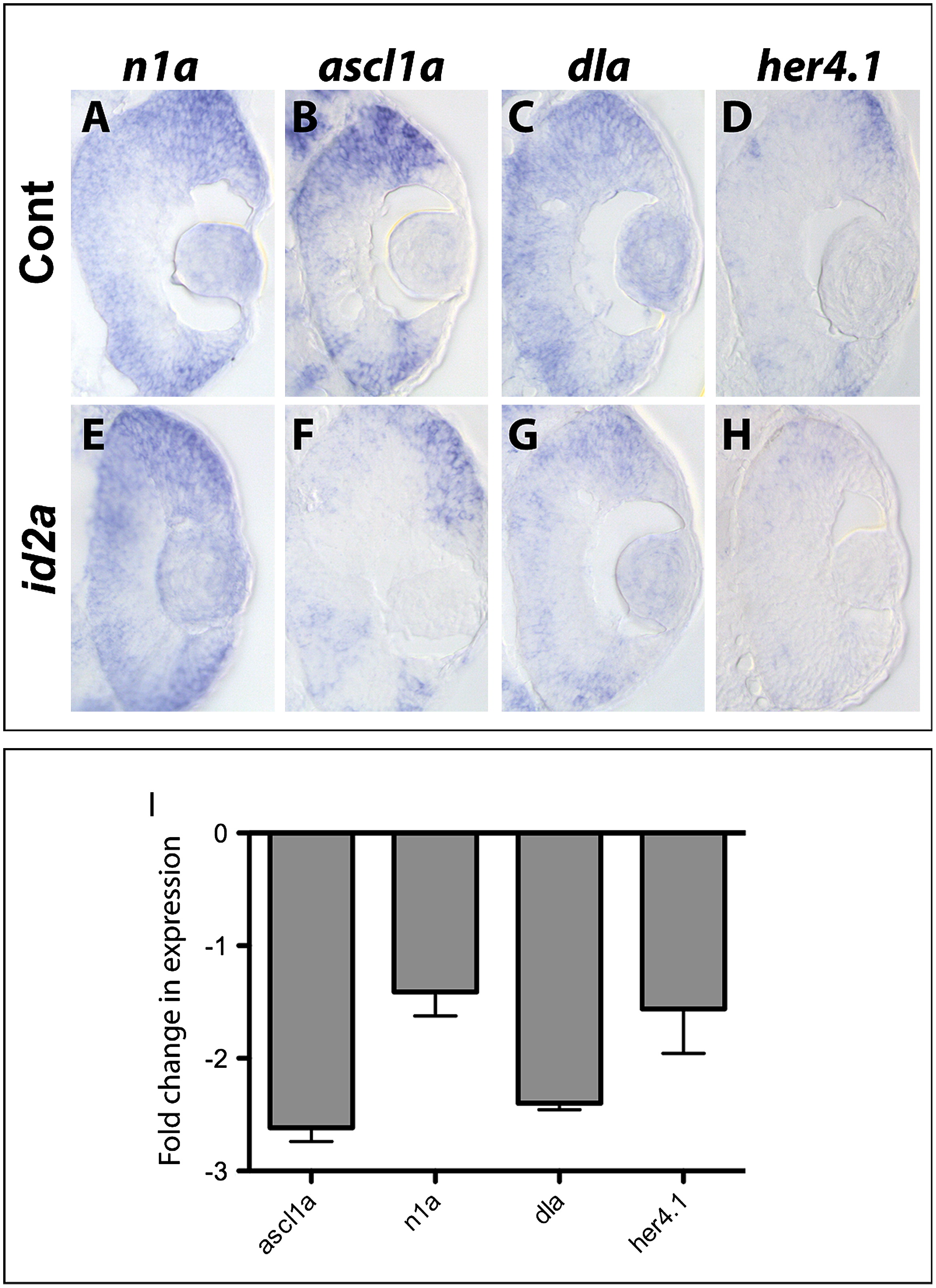
Fig. 6 Id2a-overexpression is sufficient to limit the retinal expression of Notch pathway component genes. Transverse sections reveal the expression domains for n1a, ascl1a, dla and her4.1 at 33hpf in gfp-injected control (A)–(D) and id2a-overexpressing (E)–(H) embryos following in situ hybridization (n=6 sectioned embryos/condition). (I) qRT-PCR quantification of ascl1a, n1a, dla and her4.1 levels in id2a retinas compared to gfp control retinas at 33 hpf. Transcript levels were normalized to tubulin, alpha 1 and the fold-change in expression in gfp-injected vs. id2a-injected retinae presented. Error bars represent SEM, **p <.05, n=3 biological replicates.
Reprinted from Developmental Biology, 371(2), Uribe, R.A., Kwon, T., Marcotte, E.M., and Gross, J.M., Id2a functions to limit Notch pathway activity and thereby influence the transition from proliferation to differentiation of retinoblasts during zebrafish retinogenesis, 280-292, Copyright (2012) with permission from Elsevier. Full text @ Dev. Biol.