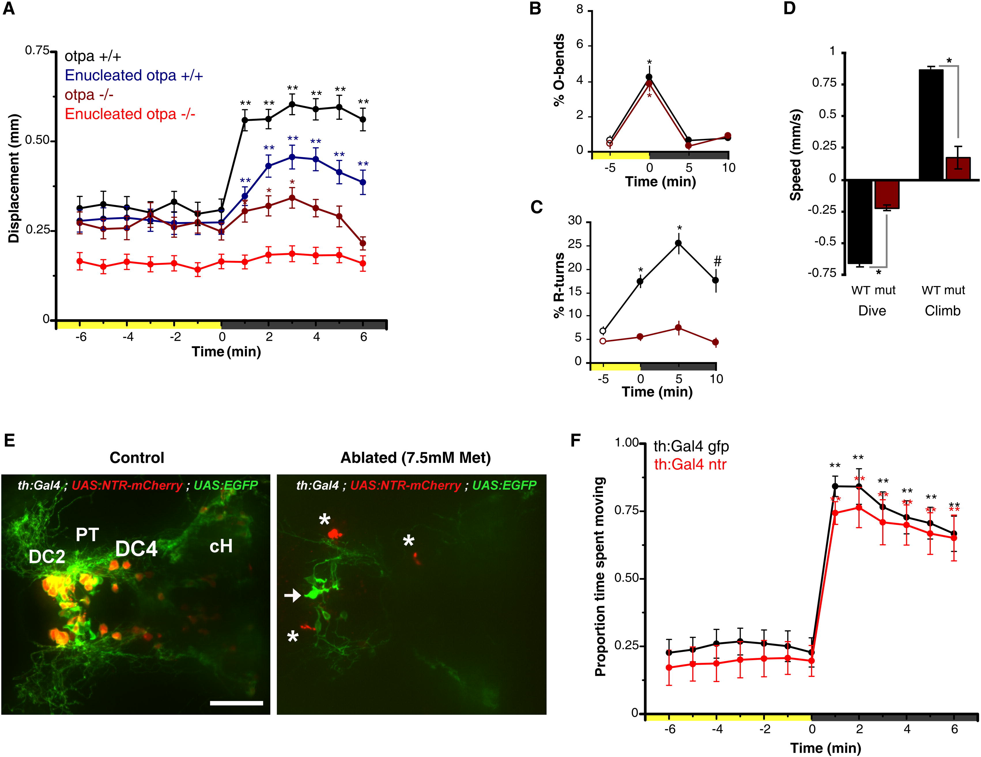
Fig. 2 Reduction of VMR in otpa Mutants and Lack of Dopaminergic Contribution(A) Locomotor activity during dark-induced VMR of intact and enucleated otpa mutants and sibling larvae. Intact mutants show a response to light extinction (repeated-measures ANOVA; F3.0,175 = 8.5, p < 0.01; n = 59 larvae) that is greatly reduced relative to controls (intact siblings: n = 47 larvae; enucleated siblings: n = 97 larvae). Without eyes, mutants lose any response to light extinction (repeated-measures ANOVA; F3.2,317 = 1.72, p = 0.16; n = 101 larvae). Data represent mean activity for the preceding minute. Color along x axis indicates light condition. Pairwise comparisons to baseline time point at 0 min: p < 0.05, p < 0.01.(B and C) Kinematic analysis of photokinesis in intact otpa mutants. Otpa mutants retain O-bend responses to light extinction (B) (repeated-measures ANOVA; F3,9 = 53.7; p < 0.001; n = 4 groups of 10 larvae) but do not show characteristic increases in R-turn initiation (C) (mutants, red; siblings, black) (F3,9 = 2.1; p = 0.17; n = 4 groups of 10 larvae). #p < 0.05, p < 0.01 for pairwise comparisons to baseline at 5 min (empty circles). Data represent the mean and SEM of observations during the first 16 s following each time point.(D) Diving and climbing speed of intact otpa mutant larvae. Compared to siblings, otpa mutants exhibit significantly reduced diving speed during and climbing speed following a 60 s dark flash (t test; dive: p < 0.001; climb: p < 0.005; n = 3 groups of 8 larvae). Data represent mean and SEM swim speed over first 20 s of dive and ascent.(E) Nitroreductase-mediated ablation of dopaminergic (DA) neurons in Tg(BACth:Gal4VP16)m1233; Tg(UAS:EGFPCAAX); Tg(UAS-E1b:NfsB-mCherry) triple-transgenic larvae. Asterisk indicates mCherry aggregates remaining from ablated cells. Arrow indicates GFP-expressing nonablated cells. The following abbreviations are used: DC2 and DC4, Otp-dependent dopaminergic groups 2 and 4; PT, posterior tuberculum; cH, caudal hypothalamus. Dorsal view is shown. Scale bar represents 50 μm.(F) VMR in DA neuron-ablated larvae. Control Tg(th:Gal4VP16); Tg(UAS:EGFPCAAX) (black line) and DA neuron-ablated Tg(th:Gal4VP16); Tg(UAS-E1b:NfsB-mCherry) (red line) larvae show similar, robust VMR following light extinction (repeated-measures ANOVA; F12, 564 = 148.29, p < 0.001; n = 48 larvae). Data represent mean and SEM activity as in (A). Pairwise comparisons to baseline time point at 5 min: p < 0.05, p < 0.01.See also Figure S2.
Image
Figure Caption
Figure Data
Acknowledgments
This image is the copyrighted work of the attributed author or publisher, and
ZFIN has permission only to display this image to its users.
Additional permissions should be obtained from the applicable author or publisher of the image.
Full text @ Curr. Biol.

