
|
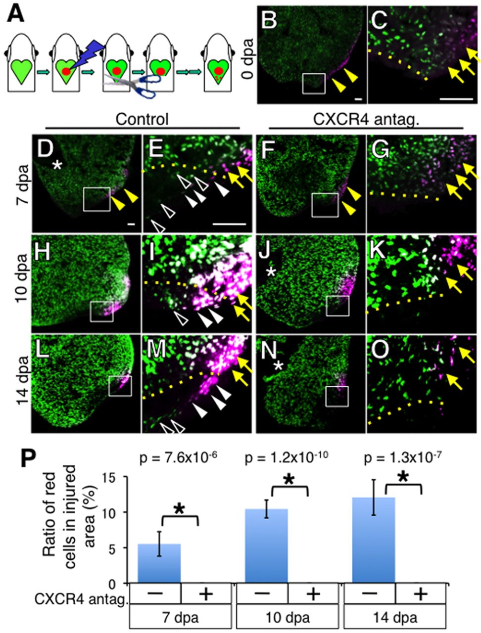
Fig. 7 Cxcr4 function is required for CM migration during heart regeneration. (A) Experimental strategy to assay CM migration during heart regeneration. After localized photoconversion of the cmlc2a-Kaede heart, hearts are exposed by enlarging the pericardiac window, and the ventricular apex is amputated. At the desired time, labeled CMs are examined by imaging analysis. (B-O) Green signal represents non-irradiated Kaede and newly synthesized Kaede after irradiation and red signal represents photoconverted Kaede. C, E, I, M, G, K and O show higher magnification images of boxed area in B, D, H, L, F, J and N, respectively. (B,C) Immediately after photoconversion and amputation, green signal was lost at the irradiated site (B, arrowheads), where red signal was detected (C, arrows). Ventricular amputation was performed in the non-irradiated area. (D,E) At 7 dpa, red signals (photoconverted CMs, white arrowheads in E) and green signals (non-photoconverted CMs, open arrowheads in E) were detected in the injury site. (H-M) At 10 dpa (H,I) and 14 dpa (L,M), red signals and green signals were also detected in the injury site. (F-O) CMs were not detected in the injury site of the CXCR4-antagonist-treated heart. All photoconverted CMs stayed outside the injury site (yellow arrows in G,K,O) by blocking Cxcr4 function. (P) Quantitation of the migrated CMs 7, 10 and 14 days after photoconversion and amputation. The vertical axis represents the percentage of photoconverted CMs in the injury site compared with the number of entire photoconverted CMs. A section at the center of the injury site was examined from each heart. Dotted lines indicate the amputation planes. The yellow arrowheads in B, D and F point to the irradiated areas. The yellow arrows in C, E, G, I, M, K and O point to red signals outside the injury site. The open arrowheads and white arrowheads point to non-photoconverted CMs and photoconverted CMs, respectively, in the regenerating area. The asterisks in D, J and N indicate the valves between the atrium and ventricle. Scale bars: 50 μm.