
Image
|
Figure Caption
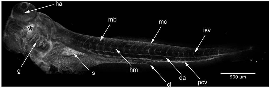
Fig. 7 HxBP labeling in a 96 hpf swimming larva.
Composite of confocal projections taken of a 96 hpf larva injected (at asterisk) with 50 μM trifunctional HxBP. Strong labeling is evident throughout the developing circulatory system, most notably in the looping vessels of the gill arches (g), the dorsal aorta (da), posterior cardinal vein (pcv), intersomitic vessels (isv), and hyaloid artery (ha). Labeling is also notable in individual migratory mesenchyme cells (mc), the protease-rich stomach (s), horizontal myoseptum (hm) and maturing craniofacial cartilages. Scale bar is 500 μm.
Acknowledgments
This image is the copyrighted work of the attributed author or publisher, and
ZFIN has permission only to display this image to its users.
Additional permissions should be obtained from the applicable author or publisher of the image.
Full text @ PLoS One