Image
Figure Caption
Fig. S7
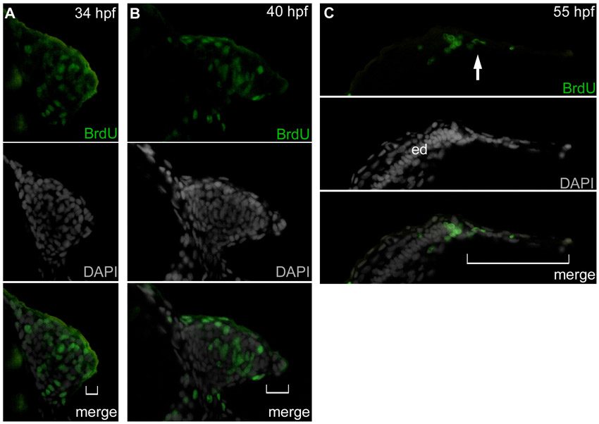
Cell proliferation in the pectoral fin bud. (A-C) BrdU-positive cells were not observed in the AER or AF (white brackets). BrdU-positive cells were mainly observed in the undifferentiated mesenchyme, in the distal periphery of the endoskeletal disc (ed), and in a few migrating mesenchymal cells (white arrow) at 34 (A), 40 (B) and 55 (C) hpf. All observations were repeated in more than eight samples, which yielded equivalent results. Upper, middle and bottom panels show immunostaining for BrdU (GFP), DAPI (gray) and merged images.
Acknowledgments
This image is the copyrighted work of the attributed author or publisher, and
ZFIN has permission only to display this image to its users.
Additional permissions should be obtained from the applicable author or publisher of the image.
Full text @ Development

