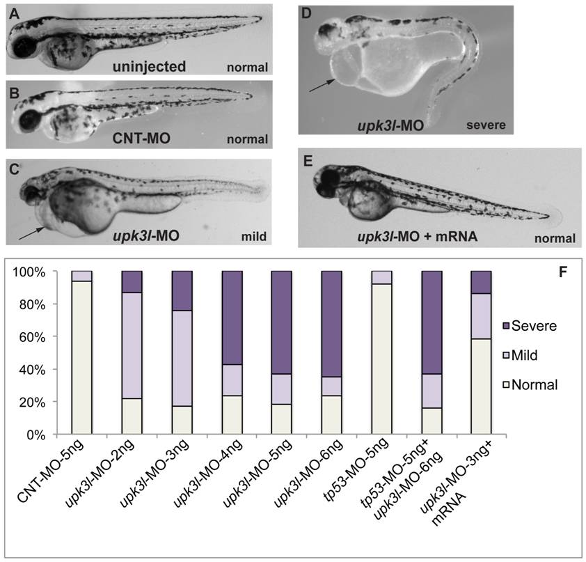
Fig. 2 Phenotypes of upk3l morphant embryos.
(A–D) Uninjected-, CNT-MO-, or upk3l-MO-injected embryos were examined under a dissecting microscope and photographed. Pericardial edema, when observed, is indicated by a black arrow. (E) Embryo injected with 3 ng upk3l-MO, 100 pg MO-resistant upk3l mRNA, and 50 pg of mCherry mRNA (to mark successful injections). Microinjection of MO-resistant mRNA alone at doses of 50-200 pg did not produce detectable phenotypic abnormalities. (F) Distribution of morphological phenotypes associated with embryos injected with 5 ng CNT-MO, 2-6 ng of upk3l-MO, tp56-MO ±upk3l-MO, or upk3l-MO/upk3l mRNA/mCherry (n = 100).

