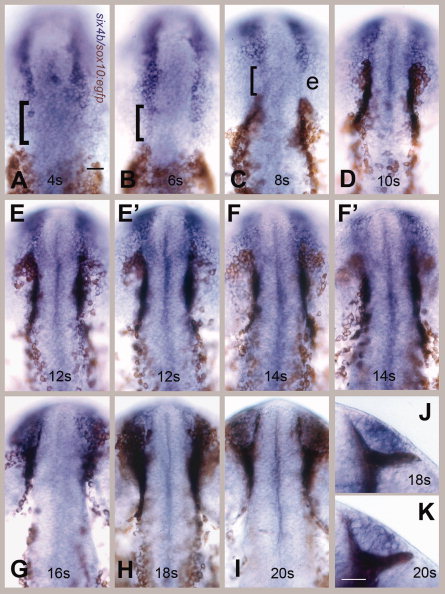
Fig. 3 six4b-expressing olfactory placode (OP) precursors do not share a common border with cranial neural crest cells (CNCCs). Visualization of OP field using six4b (in blue) and CNCCs using anti-green fluorescent protein (GFP) immunocytochemistry (in brown) in sox10:EGFP embryos. A–I: Dorsal views, rostral to the top of the page. E,E′: Two different focal planes of same embryo at 12 somites: E, dorsal OP; E′, ventral edge of the OP. F,F′: Two different focal planes at 14s (somite stage): F dorsal OP, F′ ventral edge of the OP. The neural crest cells are both dorsal and ventral to the olfactory fields at these stages. G–I: CNCCs move dorsally over the forming OP. J,K: Ventral views of the formed OP surrounded by CNCCS at 18s (J) and 20s (K), rostral to top. Scale bars = 30 μm in A (applies in A–K). Twenty embryos were examined per time point.
Image
Figure Caption
Acknowledgments
This image is the copyrighted work of the attributed author or publisher, and
ZFIN has permission only to display this image to its users.
Additional permissions should be obtained from the applicable author or publisher of the image.
Full text @ Dev. Dyn.

