Fig. 1
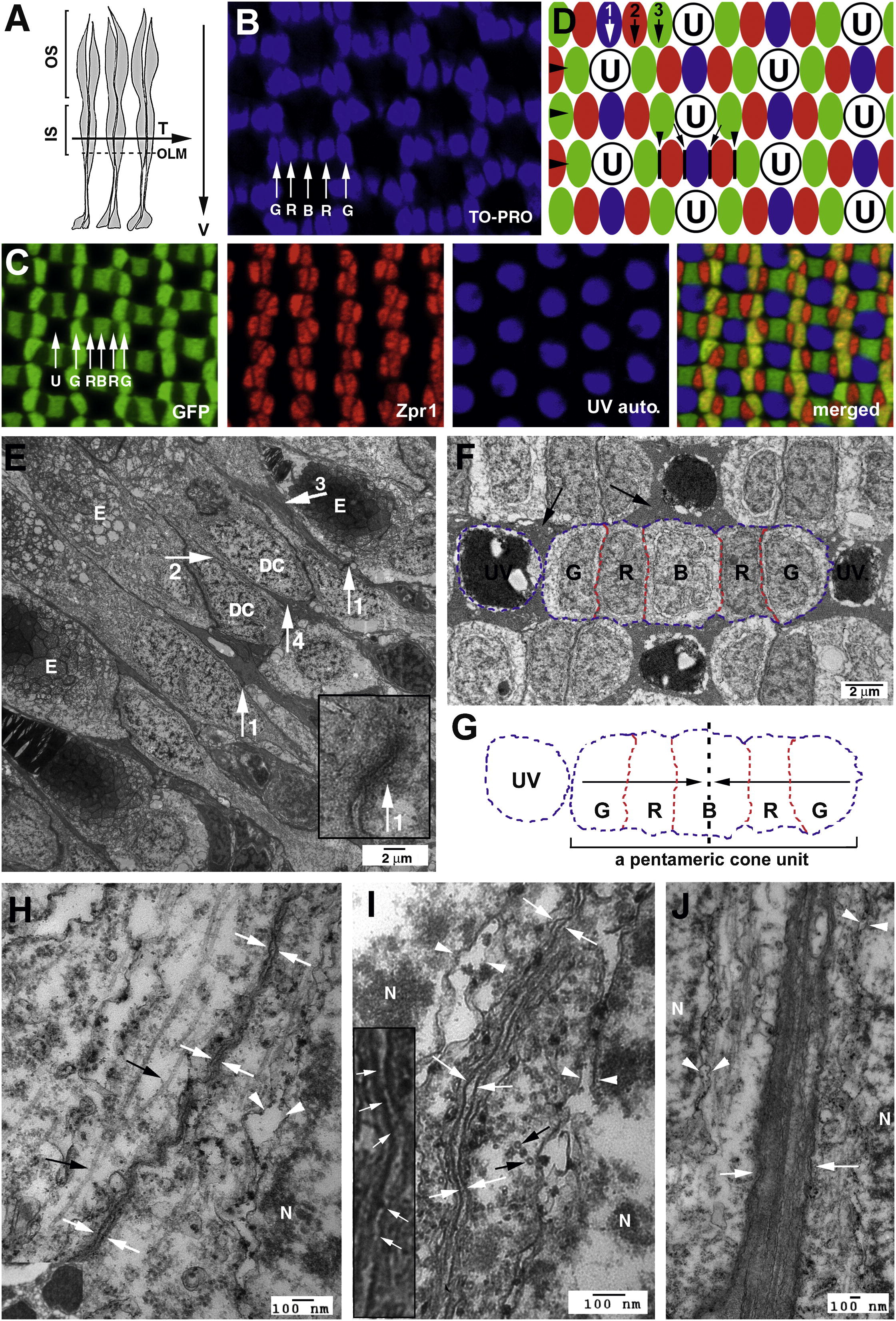
Cones Are Organized in Regular Mosaics in the Zebrafish Retina, and Their Inner Segment Cell Membranes Are Closely Juxtaposed (A) A schematic illustrates the transverse/tangential (arrow T) and vertical (arrow V) section planes of photoreceptors (gray). The outer limiting membrane (OLM; dashed line); inner segments (IS); outer segments (OS). (B) TO-PRO nuclear staining illustrates the geometric alignments of the cell nuclei of green (G), red (R), and blue (B) cones. (C) Confocal images of four types of cones in wild-type Tg(RH2-1:GFP) pt112 adult zebrafish (Zou et al., 2010), which express GFP strongly in green (G) and blue (B) cones and weakly in red (R) cones. Double cones (red) were visualized with zpr1 antibodies. UV cones (blue) were visualized by autofluorescence signals. (D) A schematic illustrates the orthogonal planar arrangements of rows (large arrowheads) and columns of cones (arrows 1, 2, and 3). The junctional interfaces (small arrowheads) between the members of double cones are parallel to the axes of cone columns. The junctional interfaces between red and blue cones are indicated with black lines and small arrows. The letter U stands for UV cones; green, red, and blue cones are color-coded in green, red, and blue, respectively. (E) Transmission electron microscopic (TEM) imaging of the inner segment vicinity of a vertically sectioned adult retina revealed the neatly aligned OLM (arrow 1s), direct juxtaposition between double cone (DC) members (arrow 2), and the bundles of long (arrow 3) and short (arrow 4) apical Müller cell processes. The ellipsoids are indicated with the letter E. The inset shows an OLM junction at a higher magnification. (F) TEM imaging of a tangentially sectioned wild-type adult retina revealed the flat junctional interfaces (red dashed lines) between members of a pentameric cone unit, which contains a blue cone (B) and two pairs of double cones (R, for red cones; G for green cones). UV cones, surrounded by clusters of Müller cell apical processes and the inner segments of rods (arrows), are round and do not establish any flat junctional interfaces with other cone types at the level of inner segments. The blue contour lines indicate the convex cell membrane regions of the cones. (G) The contours of a UV cone and a pentameric cone unit in (F) are duplicated here to better illustrate the overall shape of cones at cross sections. The dashed line and arrows illustrate the mirror-symmetrical alignment of the cone members within a pentameric cone unit. (H and I) TEM imaging of the junctions between the members of double cones (opposing white arrows), either sectioned vertically (H) or transversly (I). The inset at a higher magnification shows the fine filaments (arrows) that connect the neighboring cell membranes. (J) Müller cell apical processes are tightly bundled together (opposing white arrows). N, cell nuclei; black arrows, microtubules (H, I, and J).
Reprinted from Developmental Cell, 22(6), Zou, J., Wang, X., and Wei, X., Crb Apical Polarity Proteins Maintain Zebrafish Retinal Cone Mosaics via Intercellular Binding of Their Extracellular Domains, 1261-1274, Copyright (2012) with permission from Elsevier. Full text @ Dev. Cell

