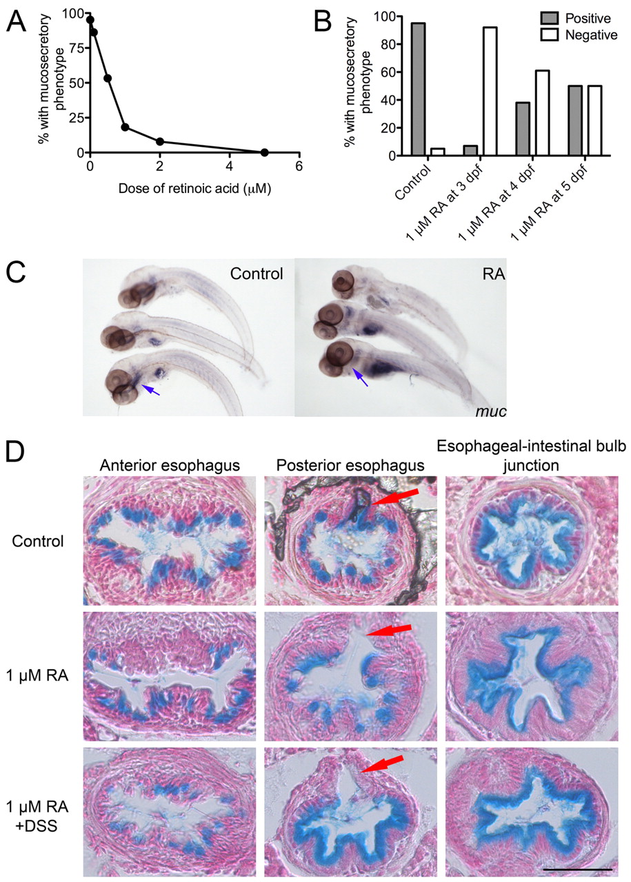
Fig. 5 Retinoic acid suppresses the DSS-induced mucosecretory phenotype. (A) Dose-response curve of the percentage of DSS-exposed larvae positive for the mucosecretory phenotype after the administration of a range of doses of RA (n≥20 per group; two biological replicates). (B) Proportion of DSS-exposed larvae positive or negative for the mucosecretory phenotype at 6 dpf after administration of RA at the day indicated. Fisher’s exact test P-values vs untreated controls: 3 dpf<0.0001, 4 dpf=0.0002, 5 dpf=0.0011; n≥20 per group, two biological replicates. (C) Whole-mount in situ hybridization detection of muc expression in the esophagus in control and RA-treated larvae. Blue arrows indicate the location of the esophagus. (D) Alcian-blue-stained esophageal sections of larvae treated with 1 μM RA and exposed to DSS. Arrows indicate the pneumatic duct. Scale bar: 50 μm.
Image
Figure Caption
Figure Data
Acknowledgments
This image is the copyrighted work of the attributed author or publisher, and
ZFIN has permission only to display this image to its users.
Additional permissions should be obtained from the applicable author or publisher of the image.
Full text @ Dis. Model. Mech.

