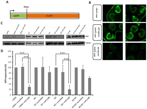
Fig. 4 GFP sensor assay.
A) The GFP gene was fused with the 3′UTR of Nrp2a that contained the putative binding sites of miR-2188. This construct was transcribed in vitro into capped mRNA prior to injection. B) Single cell embryos were injected with the GFP reporter (50 ng/μL) in the presence or absence of miR-2188 and scrambled duplexes. Fluorescence levels were observed at 24 hpf and representative embryos of each condition were photographed. Decreased fluorescence was observed in miR-2188 duplex injected embryos. C) Embryo lysates were prepared from 24 hpf embryos injected with the wild type GFP reporter or the mutated reporters in the presence or absence of miR-2188 and scrambled duplexes. Protein levels were determined by western blot analysis and β-tubulin was used as an internal control. D) Quantification of the reporter proteins (%) in the conditions tested using 3 biological replicates. Asterisks indicate conditions where GFP expression was significantly down regulated by miR-2188 relative to control conditions. miR-2188 injected embryos showed a statistically significant down regulation of GFP levels, indicating that Nrp2a 3′UTR is a true miR-2188 target. Co-injection of the M1 reporter with the miR-2188 duplex produced normal GFP levels, indicating that the BS-1 mutation abolished miR-2188 binding. On the other hand, a significant decrease in GFP expression was observed after co-injection of the M2 reporter with miR-2188, relative to the controls, indicating that BS-2 was not critical for miR-2188 binding. Data are mean +/stdev, p<0,005 (t test, unpaired), n>3. All lanes were normalized to the β-tubulin signal.

