
|
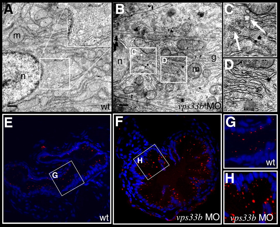
Fig. 6
vps33b knockdown disrupts intestinal vesicle transport. (A-D) Electron micrographs of enterocytes from 5 dpf wild-type (A) and vps33b morpholino injected (B-D) larvae. The apical surface is to the right of each panel. Insets are at twice the magnification of the indicated regions of cytoplasm. No vesicles can be identified within the wild-type cell cytoplasm. (C) Multiple vesicles (arrows) are seen in the cytoplasm of vps33b-deficient enterocyte. Dilated stacks of Golgi cisternae are also evident within these cells (D). g, golgi; m, mitochondria; n, nucleus. (E-H) Histological cross-sections from the anterior intestine of 5 dpf wild-type (E,G) and vps33b morpholino-injected (F,H) larvae that have ingested the styryl dye AM1-43. Nuclei stained with DAPI. (G,H) Magnified views from E and F, respectively. There was a 1.8-fold increase in the number of fluorescent vesicles in the vps33b-deficient larvae (see Table 4).