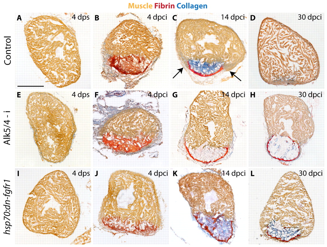
Fig. 3 Inhibition of TGFβ/Activin signaling blocks heart regeneration resulting in the infarct bulging phenotype. Acid Fuchsin Orange-G (AFOG) staining of heart sections visualizes healthy myocardium (orange), fibrin (red) and collagen (blue). (A-D) Control hearts, (E-H) Alk5/4-i-treated hearts, (I-L) hearts of transgenic fish expressing heat shock-inducible dn-fgfr1. (A,E,I) Control ventricles after sham operation (4 dps) consist of healthy myocardium. (B,F,J) At 4 dpci, similar infarcts (red) are detected in all experimental groups. (C,G,K) Hearts at 14 dpci. (C) Control hearts contain abundant fibrillar collagen in the injury area. A layer of fibrin (red) covers the margin of the infarct. New myocardium begins to invade the scar tissue (arrows). (G) Exposure to Alk5/4-i suppresses collagen deposition. No myocardial invasion is observed. (K) Inhibition of FGF signaling does not impair collagenous scar formation in comparison to control. (D,H,L) Hearts at 30 dpci. (D) In control hearts, the scar was nearly completely resolved and replaced with new muscle. (H) Alk5/4-i blocked regeneration. The infarct is surrounded by a fibrin layer but it is collagen deficient. The injury area bulges beyond the ventricular circumference. (L) Overexpression of dn-fgfr1 blocks regeneration, resulting in a collagen-rich scar. No geometrical deformation is associated with the lack of regeneration. Scale bar: 300 μm.
Image
Figure Caption
Acknowledgments
This image is the copyrighted work of the attributed author or publisher, and
ZFIN has permission only to display this image to its users.
Additional permissions should be obtained from the applicable author or publisher of the image.
Full text @ Development

