Fig. 3
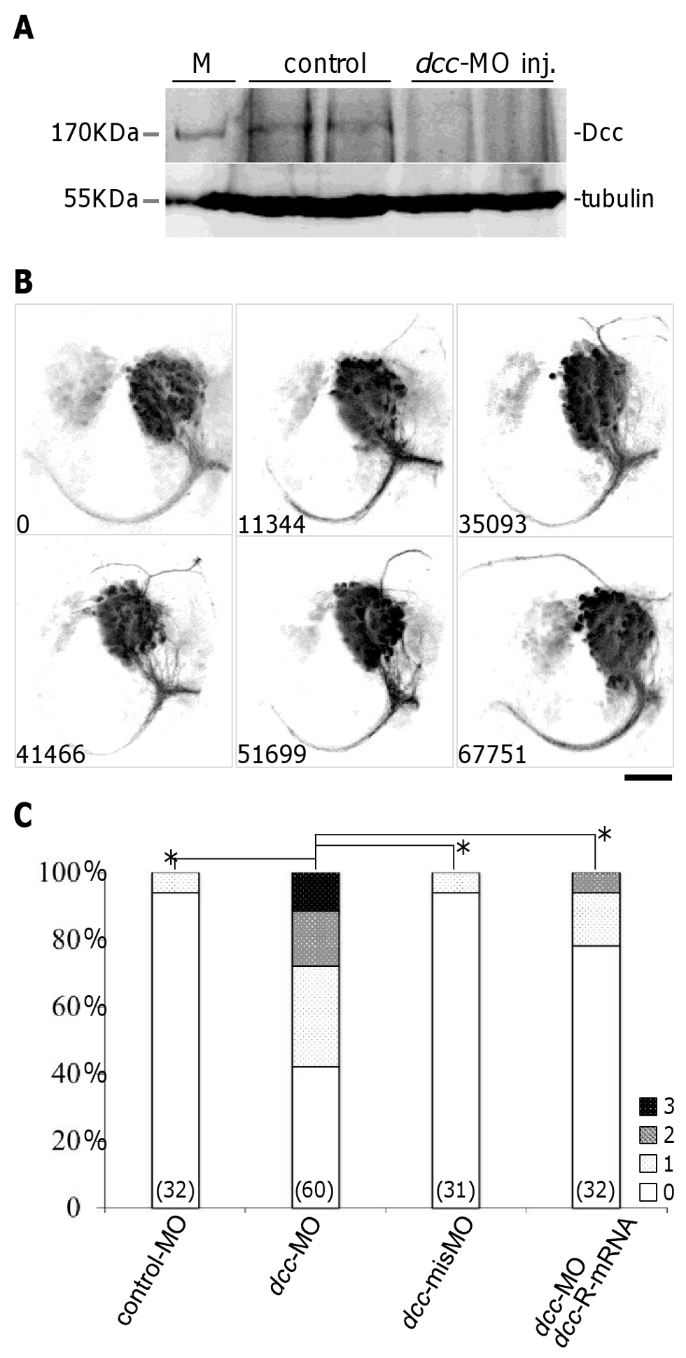
Dcc is required for correct asymmetric outgrowth of ADt neuronal axons.
(A) Dcc expression was reduced after injection of dcc translation-blocking morpholinos into zebrafish embryos. Endogenous Dcc protein was detected as a band of approximately 170 kb. Tubulin served as a loading control. M: size marker. (B) ADt neurons project axons dorsally when Dcc function is inhibited by morpholino injection. Images of live animals were acquired as in Fig. 1C. The pixel intensity value of aberrant axon is shown in the bottom left corner of each panel. Scale bar = 50 µm. (C) Quantitation of ADt neuronal axon defects. Horizontal axis shows the treatment group labels and vertical axis shows the percentage of embryos in each phenotypic category (Grade 0–3) for each treatment group. Numbers inside parentheses denote numbers of animals analyzed for each treatment group. Asterisks and brackets represent p<0.05 by Mann-Whitney U test.

