Image
Figure Caption
Fig. S3
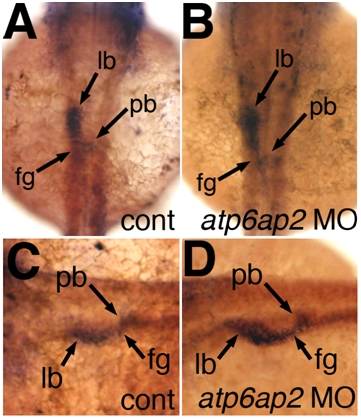
Lack of changes in the early digestive organ marker foxa3 in atp6ap2 morphants. Whole-mount in situ hybridizations of 36 hpf embryos using riboprobe directed against foxa3, which recognizes foregut derivatives. There is staining of liver bud (lb), pancreatic bud (pb) and foregut (fg) in both controls (A and C, cont) and atp6ap2 MO-injected (B and D) that appears similar. Two examples of each, control and MO-injected, are depicted.
Acknowledgments
This image is the copyrighted work of the attributed author or publisher, and
ZFIN has permission only to display this image to its users.
Additional permissions should be obtained from the applicable author or publisher of the image.
Reprinted from Developmental Biology, 365(2), Eauclaire, S.F., Cui, S., Ma, L., Matous, J., Marlow, F.L., Gupta, T., Burgess, H.A., Abrams, E.W., Kapp, L.D., Granato, M., Mullins, M.C., and Matthews, R.P., Mutations in vacuolar H(+)-ATPase subunits lead to biliary developmental defects in zebrafish, 434-444, Copyright (2012) with permission from Elsevier. Full text @ Dev. Biol.

