Fig. S7
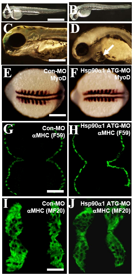
Knockdown of Hsp90a1 expression had no effect on myoblast specification and early differentiation of slow and fast myofibers. (A and B) Morphological comparison of control-MO (A) or Hsp90a1-ATG-MO (B) injected embryos at 48 hpf. The Hsp90a1 knockdown embryos appeared morphologically normal except for the lack of locomotion and muscle contraction. (C and D) Compared with control, high dose (10 ng) of Hsp90a1 ATG-MO injection induced edema in zebrafish embryos at 72 hpf (D). (E and F) In situ hybridization showing normal MyoD expression in control-MO (E) or Hsp90a1-ATG-MO (F) injected embryos at 14 hpf. (G and H) F59 antibody immunostaining on cross-sections showing expression of myosin in slow muscles of control-MO (G) or Hsp90a1-ATG-MO (H) injected embryos at 24 hpf. (I and J) MF-20 antibody staining showing expression of myosin in both fast and slow myofibers of control-MO (I) or Hsp90a1-ATG-MO (J) injected embryos at 24 hpf. (Scale bars, 250 mm in A, 50 mm in C, 150 mm in E, 75 mm in G and I.)

