
Image
|
Figure Caption
Fig. S5
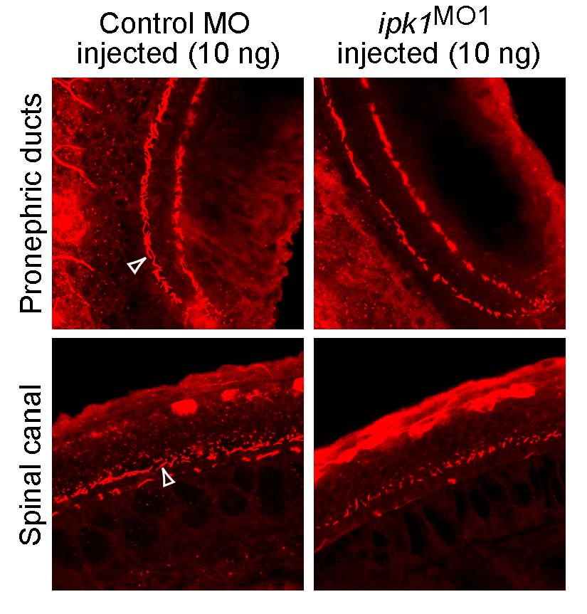
Cilia (red) in pronephric ducts (Upper) and spinal canal (Lower) of embryos at 30 hpf injected with 10 ng of either control MO (Left) or ipk1MO1 (Right). Cilia were visualized by indirect immunofluorescence with anti-acetylated tubulin antibodies and laser scanning confocal microscopy. Arrowhead indicates cilia.
Figure Data
Acknowledgments
This image is the copyrighted work of the attributed author or publisher, and
ZFIN has permission only to display this image to its users.
Additional permissions should be obtained from the applicable author or publisher of the image.
Full text @ Proc. Natl. Acad. Sci. USA