
|
Fig. 2
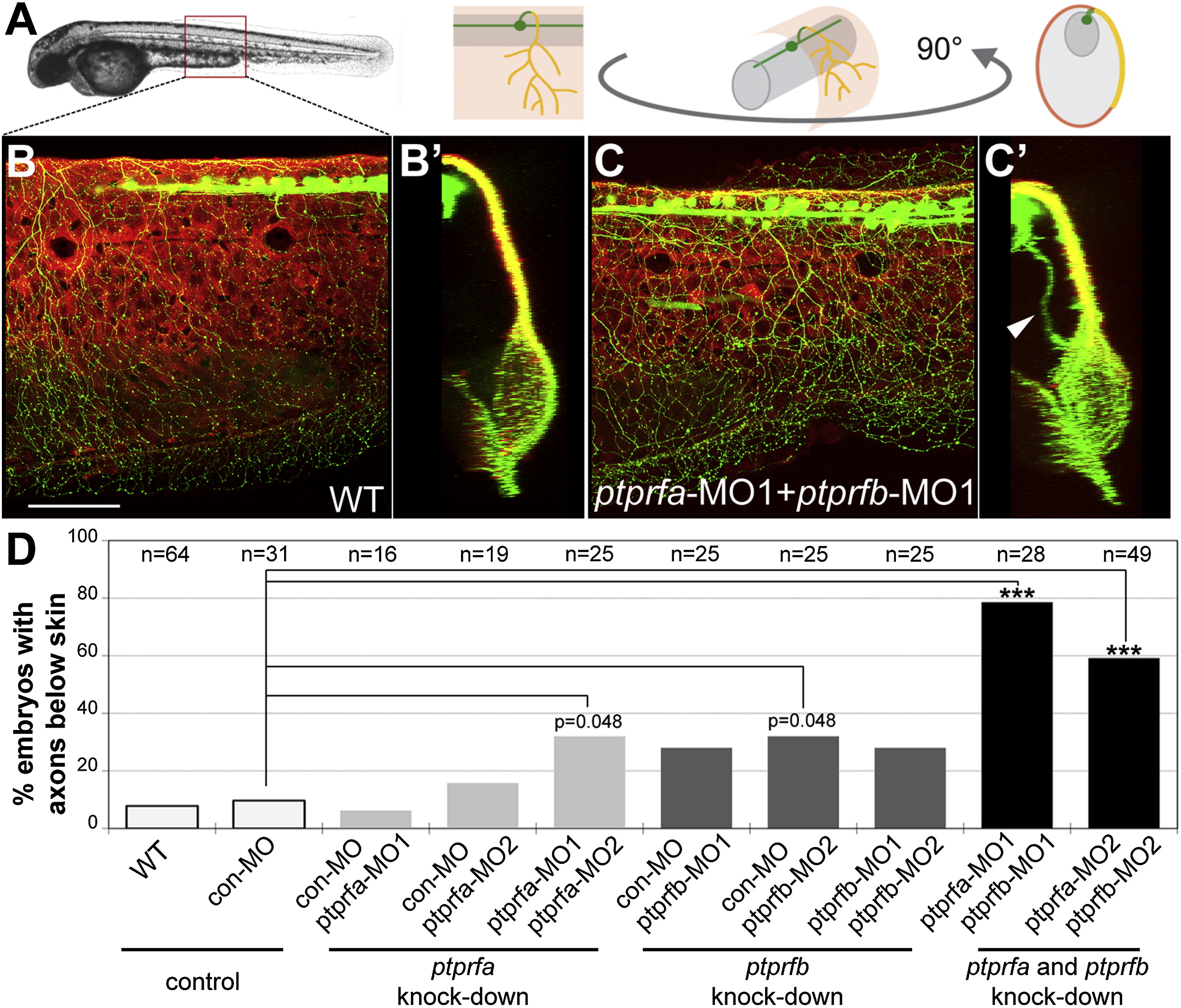
Knocking Down ptprfa and ptprfb Caused Skin Innervation Defects (A) Schematic showing the relationship between skin and axons. (B–C) Confocal projections of lateral views of WT (B) and ptprfa/ptprfb double morphant (C) embryos. 90° rotations of the images in (B) and (C) provide cross-section views of these embryos (B2–C2). Zebrafish larvae carried two transgenic reporters—sensory:GFP to visualize somatosensory neurons and krt4:dsRed to visualize the skin. Arrowhead indicates axons innervating internal tissues. Scale bar represents 100 μm. (D) Frequency of embryos with axons below the skin in a defined region of the trunk. Knocking down ptprfa or ptprfb alone caused mild skin innervation defects compared with embryos injected with an equivalent amount of control morpholino (9.7%). However, axons grew below the skin in 78.6% of ptprfa-MO1/ptprfb-MO1 double morphants and 59.2% of ptprfa-MO2/ptprfb-MO2 double morphants, which was significantly (p < 0.001) more than controls. Data was analyzed with two-sided Fisher′s exact test and computed with R (http://www.r-project.org). See Supplemental Information for details of statistical analysis. Con, control; MO, morphants; ***p < 0.001. See also Figure S2 and mmc2VIDEO and mmc3VIDEO.