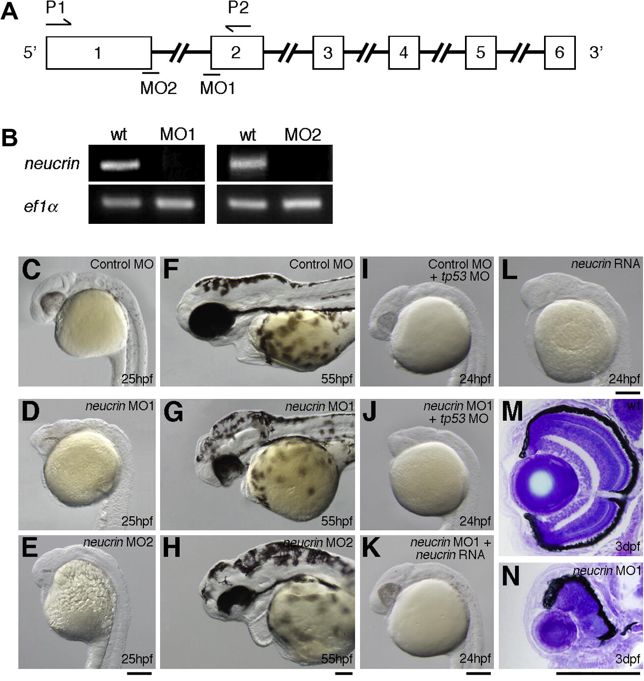
Fig. 3
Inhibition of Neucrin functions in zebrafish embryos. (A) The coding region of neucrin is divided by five introns. Open boxes and black lines indicate exons and introns, respectively. MO indicates the target position of neucrin MO. (B) neucrin cDNA was amplified from wild-type or neucrin MO-injected embryonic cDNA by RT-PCR using forward and reverse primers, the positions of which are indicated by arrows (A). ef1α cDNA was also amplified as a control. (C-H) Lateral view of control MO-injected (C, F), neucrin MO1-injected (D, G) and neucrin MO2-injected (E, H) embryos at the indicated stages are shown. MOs were injected into four central blastomeres of 16-cell embryos. (I, J) Lateral view of tp53 MO- and control MO-injected (I), and tp53 MO- and neucrin MO1-injected (J) embryos at the indicated stages are shown. tp53 MO was injected into 2- to 4-cell embryos. neucrin MO1 and control MO were injected into four central blastomeres of 16-cell embryos. (K, L) Lateral view of neucrin RNA- and neucrin MO1-injected (K), and neucrin RNA-injected (L) embryos at the indicated stages are shown. neucrin MO1 was injected into four central blastomeres of 16-cell embryos. RNA was injected into 2- to 4-cell embryos. (M, N) Transverse eye sections of wild-type (M) and neucrin MO1-injected (N) embryos at 3 dpf. Scale bar: 100 μm.
Reprinted from Mechanisms of Development, 128(11-12), Miyake, A., Nihno, S., Murakoshi, Y., Satsuka, A., Nakayama, Y., and Itoh, N., Neucrin, a novel secreted antagonist of canonical Wnt signaling, plays roles in developing neural tissues in zebrafish, 577-590, Copyright (2012) with permission from Elsevier. Full text @ Mech. Dev.

