
Image
|
Figure Caption
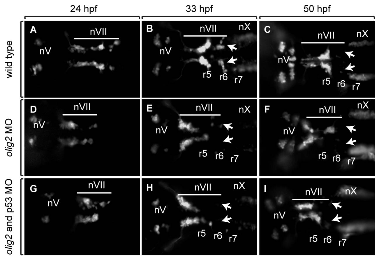
Fig. S3 Tg(isl1:gfp)rw0 reporter expression reveals that the facial motor neuron migration defect in olig2 MO-injected embryos is not affected by co-injection of p53 MO. Panels are images showing dorsal views of whole embryos with anterior to the left. A–C: Wild-type embryos. D–F: olig2 MO-injected embryos. G–I: olig2 MO-injected embryos co-injected with p53 MO. Arrows indicate posteriorly migrated facial motor neurons. Bar indicates facial motor neurons along their caudal migration. Cranial motor neurons are labeled “n.”
Acknowledgments
This image is the copyrighted work of the attributed author or publisher, and
ZFIN has permission only to display this image to its users.
Additional permissions should be obtained from the applicable author or publisher of the image.
Full text @ Dev. Dyn.