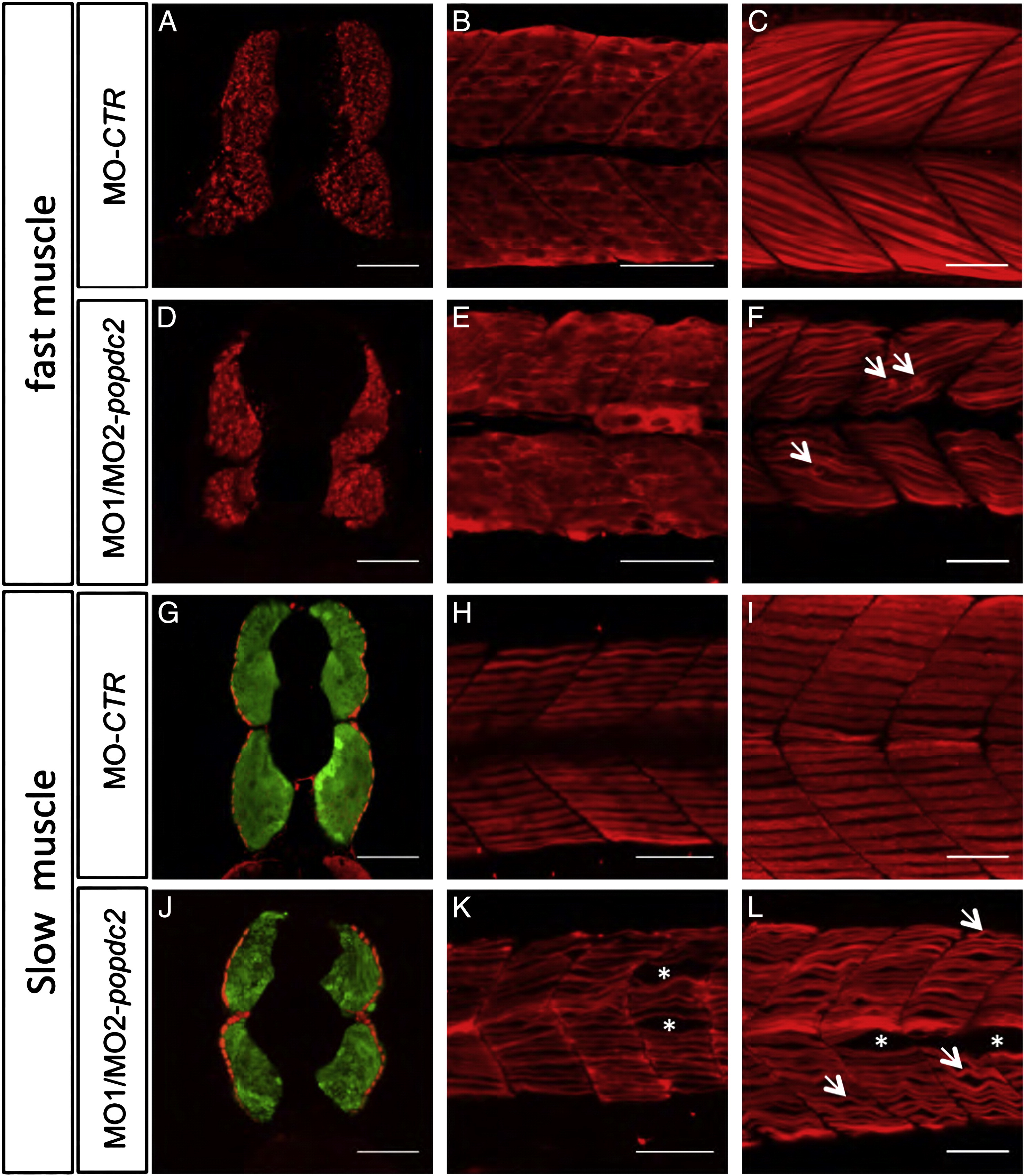
Fig. 3 Fast and slow muscle formation in popdc2 morphants. Confocal imaging of transverse sections (A,D,G,J) through the 27 hpf trunk and lateral flatmounts of (B,E,H,K) 1 dpf and (C,F,I,L) 3 dpf embryos, which were injected with (A–C,G–I) MO-control (MO-CTR) or (D–F,J–L) MO1/MO2-popdc2 morpholinos, respectively and stained (A–F) with F310 antibody to visualize fast muscle fibers, and (G–L) with F59 antibody to detect slow muscle fibers. Sections stained for slow muscle fibers (G,J) were also stained with phalloidin detecting F-actin to visualize the fast muscle compartment (green). Asterisks in (K,L) pointing to gaps in the fiber arrangement. Arrows in (F) indicate ruptured fast muscle fibers and in (L) point to slow muscle fibers with a wavy morphology. Scale bars = 50 μm.
Reprinted from Developmental Biology, 363(2), Kirchmaier, B.C., Poon, K.L., Schwerte, T., Huisken, J., Winkler, C., Jungblut, B., Stainier, D.Y., and Brand, T., The Popeye domain containing 2 (popdc2) gene in zebrafish is required for heart and skeletal muscle development, 438-450, Copyright (2012) with permission from Elsevier. Full text @ Dev. Biol.

