
|
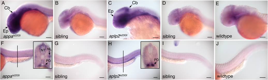
Fig. S5 rfp mRNA expression in appais22Gt and aplp2is23Gt embryos by in situ hybridization. A: RNA expression in the midbrain, cerebellum, and epiphysis in an appais22Gt embryo. F: Expression of rfp in the neural tube and pronephric ducts of an appais22Gt embryo. Line refers to position in trunk of cross-section shown in insert. Insert, rfp expression is detected in the neural tube, lateral line, and pronephric ducts. B,G: Nontransgenic sibling of the embryos in panels A and F shows no expression above background. C: RNA expression in the midbrain, cerebellum, and epiphysis in an aplp2is23Gt embryo. H: RNA expression in the pronephric ducts of an aplp2is23Gt embryo. Line refers to the position of the cross-section through the trunk shown in inset. D,I: Nontransgenic sibling of embryos shown in C and H. E,J: Wild-type embryos hybridized with rfp probe shown background level of expression. Cb, cerebellum; Ep, epiphsis; NL, neuromasts of lateral line; NT, neural tube; PD, pronephric ducts. Scale bars = 100 μm.