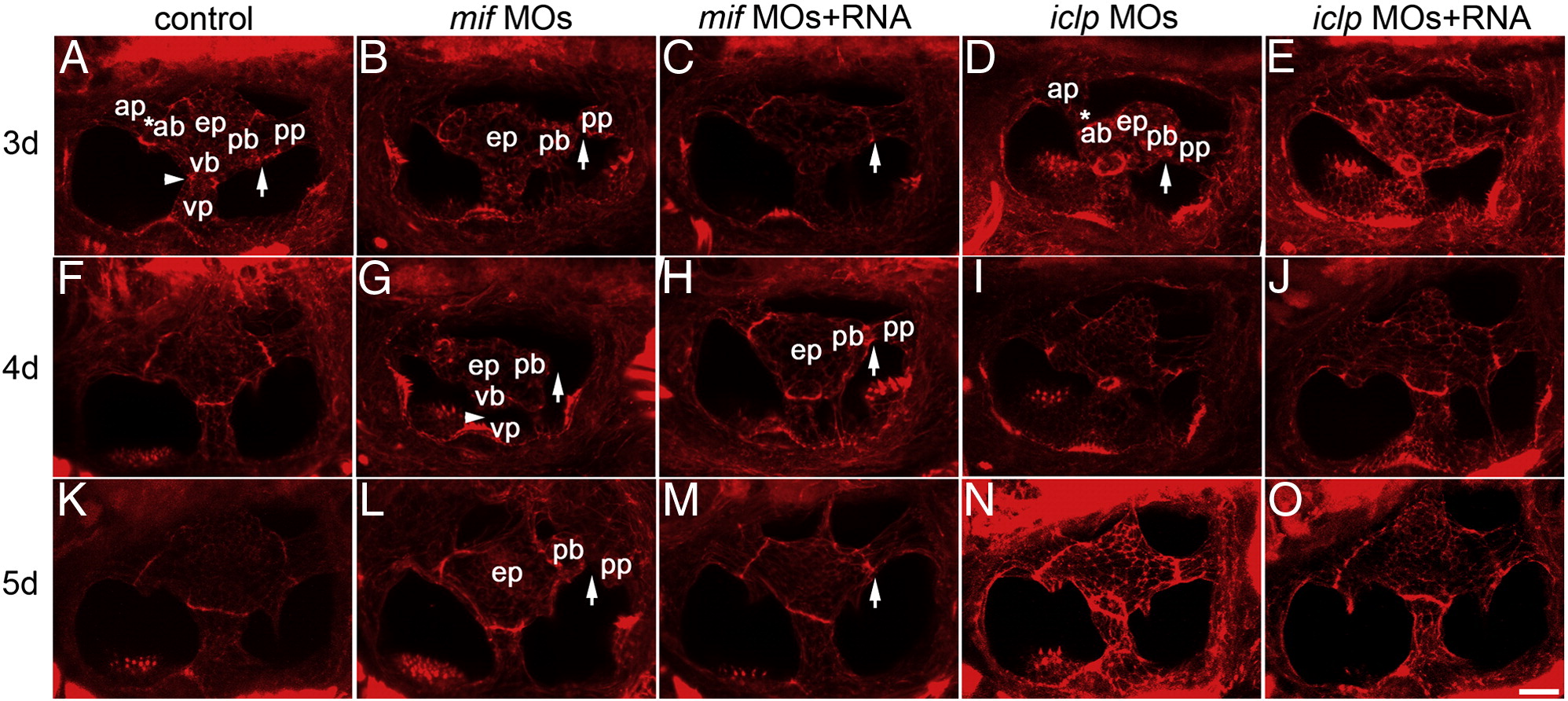
Fig. 7
Semicircular canal formation in mif and iclp morphants. (A–O) phalloidin staining of epithelial pillars (ep), which form hubs of the developing SCC. (A–E) 3 dpf; (F–J) 4 dpf; (K–O) 5 dpf larvae. (A, F, K) control; (B, G, L) mif morphants; (C, H, M) mif morphants with capped mif RNAs; (D, I, N) iclp morphants; (E, J, O) iclp morphants with capped iclp RNAs. Anterior is to the left. Arrows: junction of the posterior bulge (pb) and posterior protrusion (pp). Arrowheads: junction between the ventral bulge (vb) and ventral protrusion (vp). Stars: junction of the anterior protrusion (ap) and the anterior bulge (ab). In mif morphants, a gap between the pb and the pp was observed, indicating fusion failure. Scale bar: 25 μm. n = 11 for the 3 dpf control, 12 for the 3 dpf mif morphants, 4 for the 3 dpf mif MOs + RNA. Among the 12 mif morphants, 9 had defects in SCC formation (75%). n for 4 dpf is 7 for the controls, 6 for the mif morphants, 6 for the mif MOs + RNA, 11 for the iclp morphants, 5 for the iclp MOs + RNA. Four out of 6 (67%) mif morphants had SCC defects. For 5 dpf larvae, n = 6 for control, 7 for mif morphants, 7 for mif MOs + RNA, 5 for iclp morphants, 6 for iclp MOs + RNA. Eighty six percent (6 out of 7) mif morphants had SCC defects.
Reprinted from Developmental Biology, 363(1), Shen, Y.C., Thompson, D.L., Kuah, M.K., Wong, K.L., Wu, K.L., Linn, S.A., Jewett, E.M., Shu-Chien, A.C., and Barald, K.F., The cytokine macrophage migration inhibitory factor (MIF) acts as a neurotrophin in the developing inner ear of the zebrafish, Danio rerio, 84-94, Copyright (2012) with permission from Elsevier. Full text @ Dev. Biol.

