
|
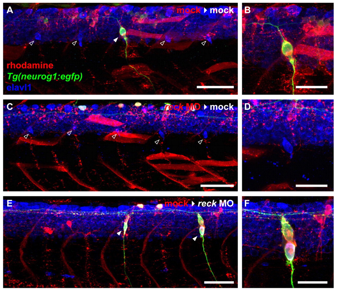
Fig. 6
DRG defects from reck depletion are cell-autonomous. (A) Mock-injected zebrafish embryo (3 dpf) implanted with mock-injected cells. Four host-derived DRG are visible (empty arrowheads) flanking one donor-derived DRG (filled arrowhead). Scale bar: 50 μm. (B) High magnification image of the donor-derived DRG shown in A. Scale bar: 25 μm. (C) Mock-injected embryo (3 dpf) implanted with MO-injected cells. Five host-derived DRG are visible (empty arrowheads), but donor cells never give rise to DRG. Scale bar: 50 μm. (D) High magnification image of one of the host-derived DRG shown in C. Scale bar: 25 μm. (E) MO-injected embryo (3 dpf) implanted with mock-injected cells. Only donor-derived DRG (filled arrowheads) are present. Scale bar: 50 μm. (F) High magnification image of one of the donor-derived DRG shown in E. Scale bar: 25 μm.