
|
Fig. S2
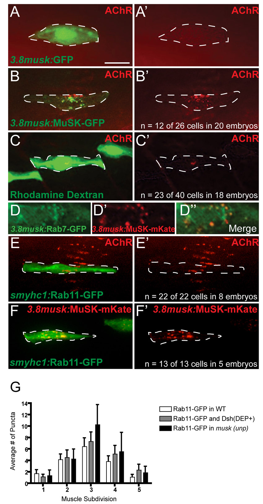
3.8musk:MuSK-GFP rescues AChR clustering to the same degree as transplanting wild-type cells into musk mutants, and distribution of Rab11-GFP and Rab7-GFP in muscle cells. (A,A2) Control expression of 3.8musk:GFP (green) in musk(unp) mutants fails to rescue AChR pre-patterning (bungarotoxin, red) (merged channels in A and red alone in A2). (B,B2) Expression of 3.8musk:MuSK-GFP in unplugged mutants at the 20-somite stage rescues AChR clusters (bungarotoxin, red) cell-autonomously in 47% of embryos (n=12 of 26 muscle cells in 20 embryos; merged channels in B, red alone in B2). (C,C2) 20-somite-stage wild-type derived adaxial muscle cell labeled with rhodamine dextran (green; transplanted at blastula stage) in an otherwise musk(unp) mutant embryo rescues AChR clusters (bungarotoxin, red) in 57% of embryos (n=23 of 40 cells in 18 embryos; merged channels in C, red alone in C2). (D-D22) Magnified view of the center of a 20-somite-stage muscle fiber showing weak colocalization of Rab7-GFP (green, green channel alone in D) and MuSK-mKate (red, red channel alone in D2) under the promoter (merged channels in D22). (E-F2) High level expression of rab11-GFP (green) via expression of smyhc1:rab11-GFP in individual, wild-type 20-somite-stage muscle cells does not alter AChR localization (bungarotoxin, red; n=22 of 22 cells in eight embryos; E,E2) or MuSK-mKate localization (n=13 of 13 cells in five embryos; F-F2). (G) Quantification of the distribution of Rab11-GFP puncta in wild-type muscle cells, musk mutant muscle cells or in cells co-expressing Dsh(DEP+)-myc. Scale bar: 10 μm.