
|
Fig. S4
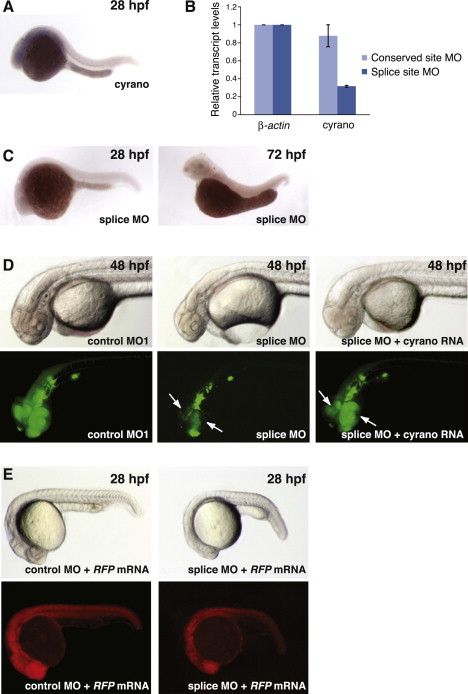
Characterization of linc-oip5 (cyrano) and NeuroD Expression in Zebrafish Embryos, Related to Figure 5 (A) In situ hybridization showing cyrano expression in the brain and notochord of zebrafish embryos at 28 hpf. (B) Reduced cyrano levels in embryos injected with the cyrano splice-site MO measured by qRT-PCR. (C) In situ hybridization of cyrano in embryos injected with the cyrano splice-site MO. (D) Embryos at 48 hpf that had been injected with the indicated reagents. Bottom panel shows subsets of neurons expressing GFP driven by the neurod promoter. Arrows point at NeuroD-positive neurons in the retina and tectum at 48 hpf. (E) Embryo coinjected with mock RNA (RFP) and the indicated MOs. Bottom panel shows RFP expression.
Reprinted from Cell, 147(7), Ulitsky, I., Shkumatava, A., Jan, C.H., Sive, H., and Bartel, D.P., Conserved Function of lincRNAs in Vertebrate Embryonic Development despite Rapid Sequence Evolution, 1537-1550, Copyright (2011) with permission from Elsevier. Full text @ Cell