
|
Fig. 7
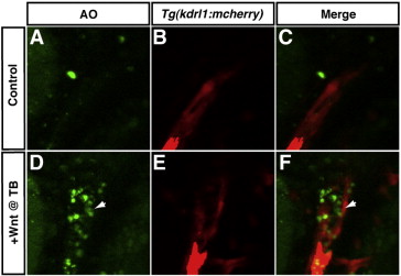
Increased Wnt signaling at the TB stage induces cell death in the anterior LPM. (A–C) Representative HCSE. (D–F) Representative GFP + embryo after increasing Wnt signaling at the TB stage. Hemizygous Tg(hsp70l:wnt8a-GFP)w34 fish were crossed to hemizygous Tg(kdrl:mCherry)ci5 fish to examine the position of apoptotic cells relative to endothelial cells in the LPM. Images are from a single optical plane taken with a Zeiss Axio Imager and ApoTome. In the transgenic embryo with increased Wnt signaling at the TB stage, there are AO positive apoptotic cells adjacent to the endothelial cells (arrows in D and F), while the HCSE does not have a significant cluster of apoptotic cells in the same region of the anterior LPM (A and C). Images are dorso-lateral views with anterior to the top and dorsal to the left.
Reprinted from Developmental Biology, 361(2), Dohn, T.E., and Waxman, J.S., Distinct phases of Wnt/β-catenin signaling direct cardiomyocyte formation in zebrafish, 364-76, Copyright (2012) with permission from Elsevier. Full text @ Dev. Biol.