
|
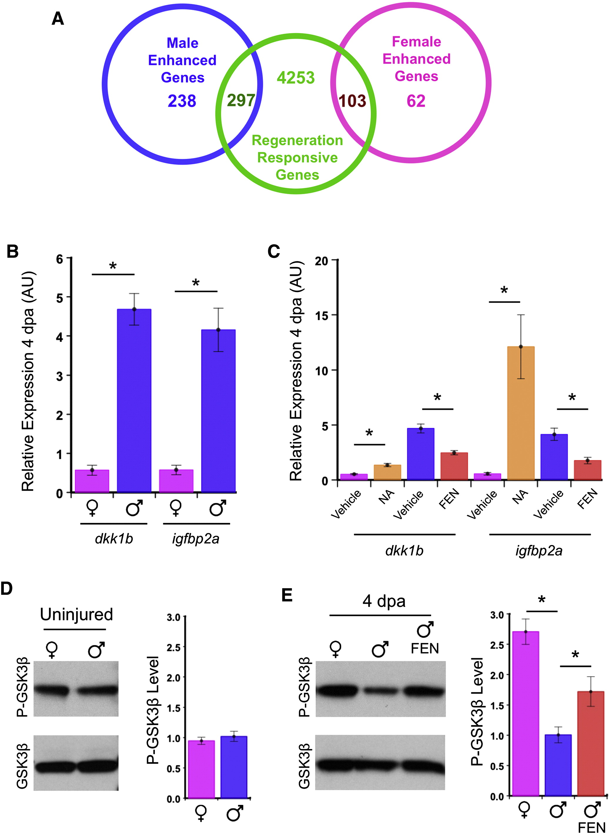
Fig. 3 Sexually Dimorphic Gene Expression and GSK3β Regulation in Regenerating Fins
(A) Venn diagram displaying numbers of genes influenced by sex or regeneration (4 dpa) and their overlaps.
(B) Increased expression of dkk1b and igfbp2a in the anterior regions of regenerating male pectoral fins compared with females (n = 3, mean ± SEM). *p < 0.05 by Student′s t test, normalized to β-actin1. AU indicates arbitrary units.
(C) NA treatment of females increased dkk1b and igfbp2a levels during anterior pectoral fin regeneration, whereas FEN treatment of males reduced their expression (n = 3, mean ± SEM). *p < 0.05 by Student′s t test, normalized to β-actin1.
(D) Uninjured male and female zebrafish pectoral fins have similar levels of inactive P-GSK3β. Values are normalized to the male P-GSK3β level (n = 3, mean ± SEM).
(E) Upon injury and regeneration, females increase P-GSK3β levels. Male P-GSK3β levels appear stable during regeneration but can be enhanced by androgen receptor antagonism. Values are normalized to the vehicle-treated male P-GSK3β level (n = 4, mean ± SEM). *p < 0.05 by Student′s t test.