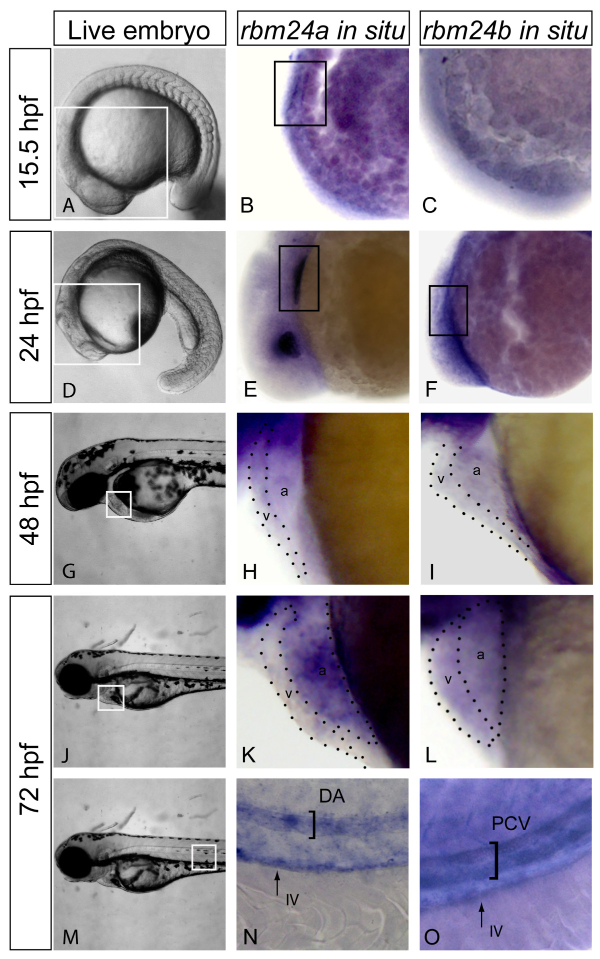
Fig. 1 rbm24a and rbm24b display cardiovascular expression during embryogenesis. Expression of rbm24 transcripts was evaluated in uninjected embryos fixed at 15.5, 24, 48 and 72 hpf. Live embryos 15.5 hpf with area of interest for rbm24 in situ boxed in white (A). Lateral heart views of rbm24a and rbm24b in situs on uninjected 15.5 hpf embryos showed expression linearly organized myocardial precursor cells for rbm24a (black box, B) but not rbm24b (C). Live embryos 24 hpf with area of interest for rbm24 in situ boxed in white (D). Lateral heart views of rbm24a and rbm24b in situs showed expression in the developing heart tube at 24 hpf (black box, E, F) with lens expression for rbm24a alone. 48 hpf live embryo showing the heart boxed in white (G). Lateral zoom of the heart showed rbm24a and rbm24b were expressed in the ventricle (v) and atrium (a) of the looped heart at 48 hpf (H, I). Live 72 hpf embryos with the heart boxed in white (J). Expression of both rbm24 transcripts was detected in the heart at 72 hpf (K, L). Live image of a 72 hpf embryo with the area of interest for vascular expression boxed in white (M). Expression of both rbm24 transcripts was detected in the trunk vasculature with differing expression patterns. rbm24a shows arterial expression in the DA (N) while rbm24b shows venous expression in the PCV (O) with both being expressed in the IV. DA, dorsal aorta; PCV, posterior caudal vein; IV, intestinal vasculature.
Image
Figure Caption
Figure Data
Acknowledgments
This image is the copyrighted work of the attributed author or publisher, and
ZFIN has permission only to display this image to its users.
Additional permissions should be obtained from the applicable author or publisher of the image.
Full text @ BMC Dev. Biol.

