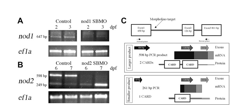
Fig. S3 Confirmation of Nod knockdown in zebrafish larvae. RT-PCR detection of (A) nod1 and (B) nod2 transcripts following MO-mediated depletion from 2 and 3 dpf (A) and 6 and 7 dpf (B) larvae. Ef1a was used as a control. (C) In silico analysis of predicted proteins derived from zebrafish Nod2 splice variants. The genomic structure with morpholino target and primer binding sites is shown at top. Exons amplified by RT-PCR are depicted as individual arrows and as a continuous amplicon of mRNA. Predicted protein structure is illustrated with shading indicating the contribution of each exon to the predicted domains.
Image
Figure Caption
Acknowledgments
This image is the copyrighted work of the attributed author or publisher, and
ZFIN has permission only to display this image to its users.
Additional permissions should be obtained from the applicable author or publisher of the image.
Full text @ Dis. Model. Mech.

