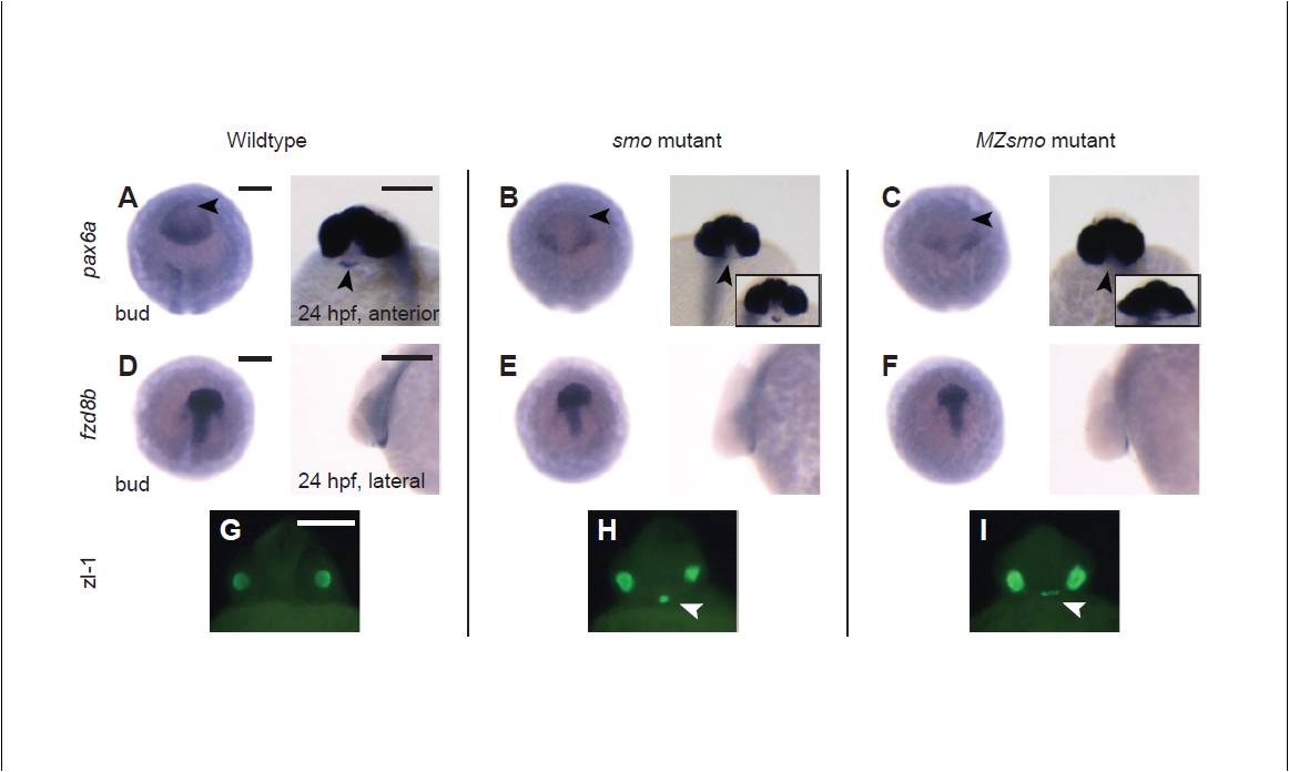
Fig. S1 Maternal smo promotes patterning of the adenohypophyseal placode. (A-C) Wild-type, smo and MZsmo embryos were stained for pax6a transcripts, which label the anterior boundary of the anterior neural plate at bud stage (10 hpf) and the adenohypophysis by 24 hpf (arrowheads). Insets in B and C depict embryos with less severe anterior pituitary defects within the group. (D-F) Riboprobes against fzd8b were used to mark the anterior neural plate in bud-stage embryos and the neurohypophysis in 24-hpf embryos. (G-I) Immunofluorescence using the zl-1 antibody marks normal and ectopic (arrowheads) lens fiber tissue, which is slightly disorganized in MZsmo embryos. Embryo orientations: A-F (bud stage), dorsal view of anterior pole and anterior to the top; A-C (24 hpf), ventroanterior view and anterior to the top; D-F (24 hpf), lateral view and anterior to the left; G-I, ventroanterior view and anterior to the top. Scale bars: 200 μm in A-F; 100 μm in G-I.
Image
Figure Caption
Acknowledgments
This image is the copyrighted work of the attributed author or publisher, and
ZFIN has permission only to display this image to its users.
Additional permissions should be obtained from the applicable author or publisher of the image.
Full text @ Development

