Fig. 4
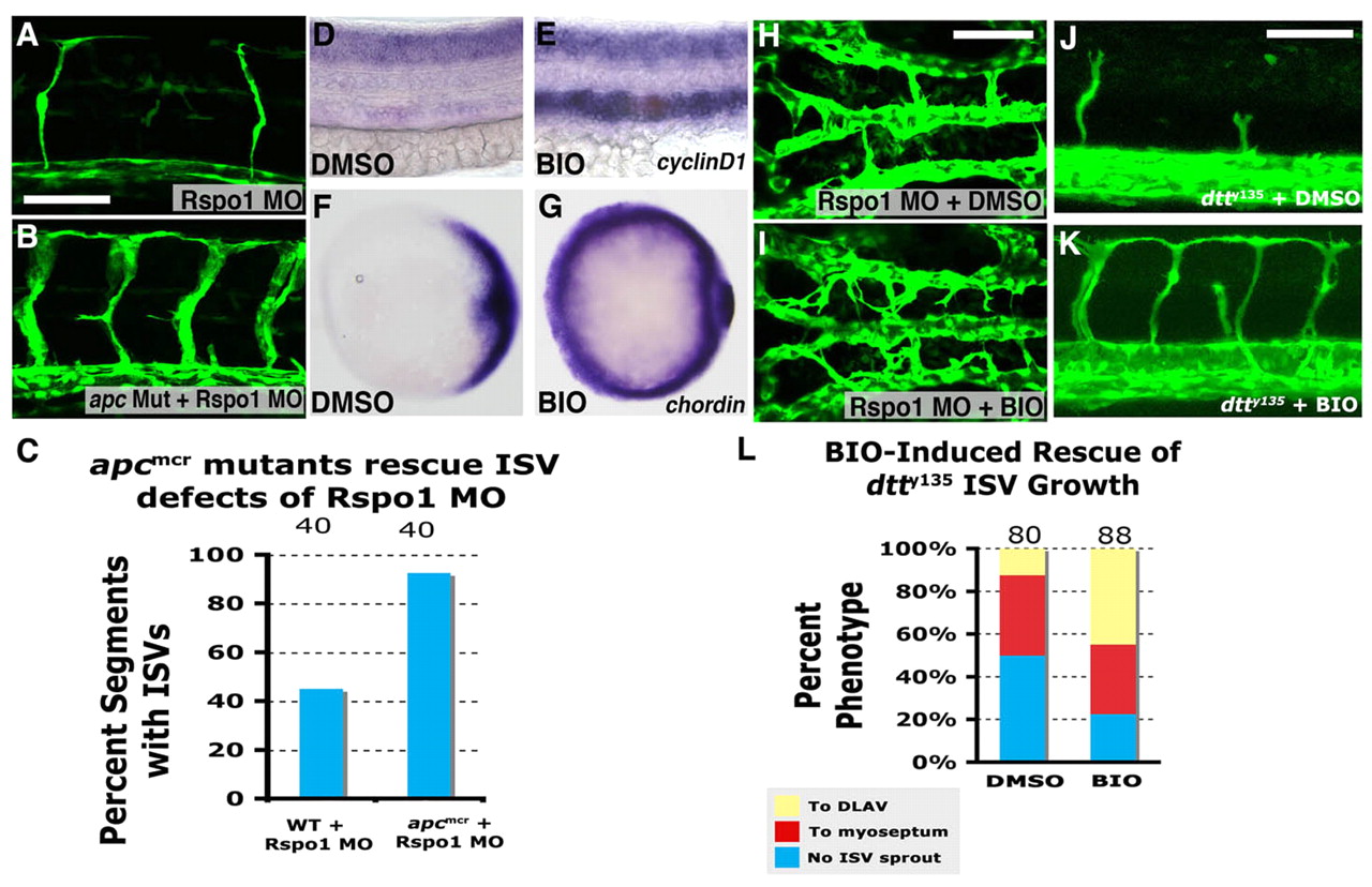
Upregulation of the Wnt/β-catenin pathway promotes angiogenesis. (A,B) Confocal images of trunk vessels in 48 hpf Rspo1 MO-injected wild-type (+/+) or heterozygous (apcmcr/+) embryo with ISV defects (A) or homozygous (apcmcr/apcmcr) mutant (B) with relatively normal ISV growth. (C) Quantitation of ISV phenotype of Rspo1 MO-injected wild type (+/+) or heterozygous (apcmcr/+) embryo and homozygous (apcmcr/apcmcr) mutant. (D,E) In situ hybridization of the trunks of 26 hpf wild-type zebrafish embryos treated from 16 hpf to 24 hpf with either carrier DMSO (D) or BIO (E), probed for cyclin D1. (F,G) In situ hybridization of the trunks of 6 hpf wild-type zebrafish embryos treated from 2.5 hpf to 6 hpf with either carrier DMSO (F) or BIO (G), probed for chordin. (H,I) Confocal images of hindbrain vessels in 48 hpf Rspo1 MO-injected embryos treated with carrier DMSO (H) or BIO (I). (J,K) Confocal images of trunk vessels in 40 hpf dtty135 mutant embryos treated with carrier DMSO (J) or BIO (K) from 16 hpf. (L) Quantitation of the intersegmental vessel (ISV) phenotypes of 40 hpf Tg(fli:EGFP)y1 dtty135 mutants treated from 16-40 hpf with either DMSO carrier or BIO. Bars show the percentages of ISV that have failed to sprout (blue), ISV that have grown only up to the horizontal myoseptum half-way up the trunk (red) and ISV that have grown all the way to the dorsal trunk to form the DLAV (yellow). The number of segments counted is shown above each bar. Scale bars: 50 μm in A,H,J.

