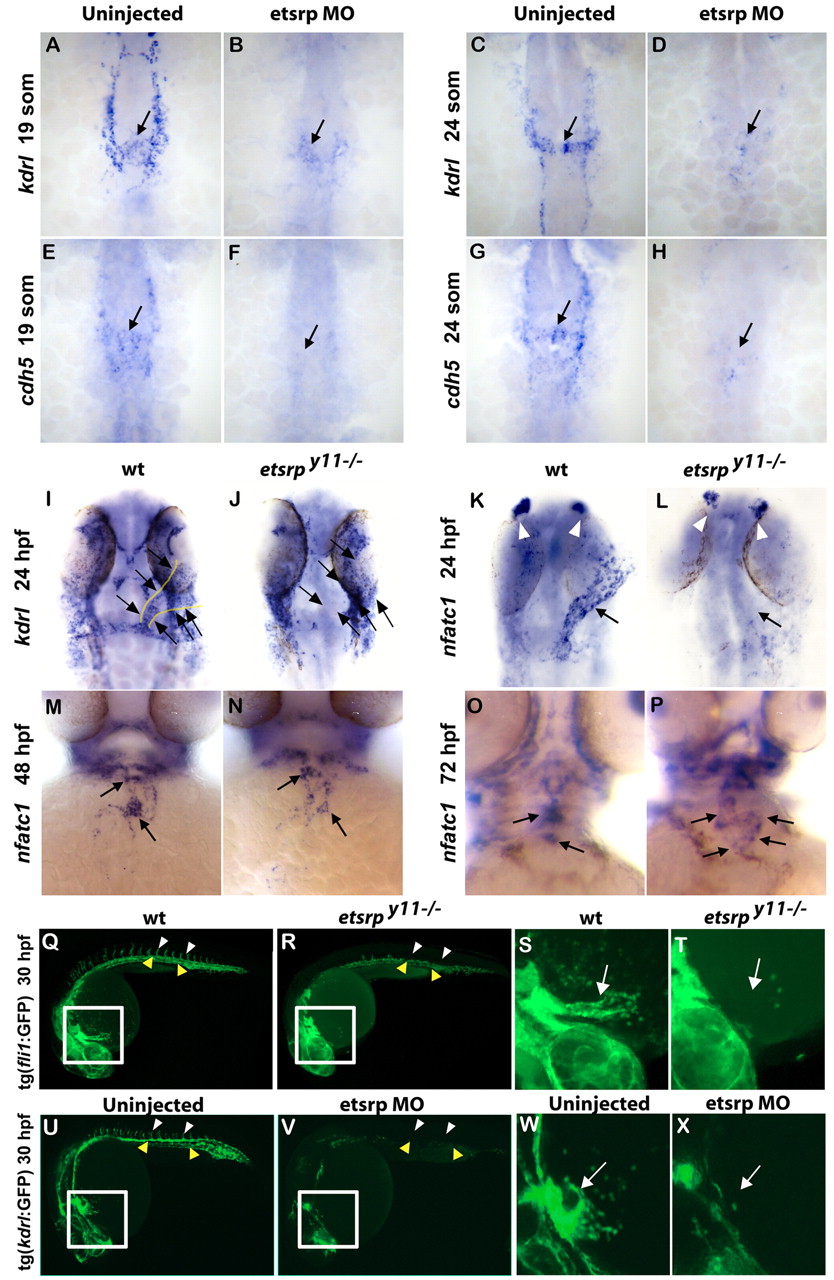
Fig. 1 Etsrp function is required for endocardium formation. (A-H) Morpholino knockdown of Etsrp results in the loss of early endocardial precursors (arrows) that migrate to the midline, as analyzed by in situ hybridization for endothelial/endocardial markers kdrl (A-D) and cdh5 (E-H) at the 19-somite (A,B,E,F) and 24-somite (C,D,G,H) stages. (I-L) etsrpy11–/– mutants lack kdrl (I,J) and nfatc1 (K,L) expression within the endocardial tube (arrows) at 24 hpf. Normal kdrl expression within the endocardium is outlined in yellow (I). nfatc1 expression in olfactory placodes is not affected (white arrowheads). (M-P) At 48 hpf (M,N) and 72 hpf (O,P) stages, nfatc1 expression in wild-type sibling embryos (M,O) is concentrated at the atrial/ventricular boundary (lower arrows) and the ventricular/outflow track boundary (upper arrows), but is sparse and diffuse in etsrpy11–/– mutants (N,P). (A-L) Ventral flat-mount view, anterior is upwards; (M-P) Ventral whole-mount view. (Q-X) Tg(fli1:GFP) (Q-T) and Tg(kdrl:GFP) (U-X) lines reveal loss of endocardial GFP in etsrpy11–/– mutants (R,T) and Etsrp morphants (V,X) at 30 hpf (insets in Q,R,U,V are shown a higher magnification in S,T,W,X, respectively). As expected, loss of Etsrp function results in the absence of intersegmental vessels (white arrowheads) and downregulation of kdrl:GFP and fli1:GFP in the axial vessels (yellow arrowheads). Lateral whole-mount view, anterior is towards the left. Arrows indicate endocardial tube.
Image
Figure Caption
Acknowledgments
This image is the copyrighted work of the attributed author or publisher, and
ZFIN has permission only to display this image to its users.
Additional permissions should be obtained from the applicable author or publisher of the image.
Full text @ Development

